For companion animals, the cutaneous system is not merely an aesthetic coat; it is the largest organ of the body, functioning as a primary immunological barrier, thermoregulatory network, and sensory interface. Among domestic felines (Felis catus), dermatological abnormalities frequently serve as external markers for systemic metabolic shifts, environmental stress, or infectious pathogens.
One of the most ubiquitous yet routinely misdiagnosed clinical signs brought to veterinary attention is the accumulation of visible epidermal flakes—commonly referred to as feline dandruff or ketombe kucing.
When a cat owner observes fine white particulates scattered along their pet’s dorsal line, the immediate instinct is to view it as a minor cosmetic nuisance resulting from exploration. However, from a clinical perspective, chronic desquamation represents an active disruption in the delicate homeostatic balance of epidermal cellular turnover, sebaceous gland secretion, and systemic hydration.
This comprehensive manual delivers an exhaustive analysis of feline dermatological health. It maps out the cellular mechanics of keratinization, explores the differentiating diagnostic matrices between benign flaking and contagious zoonotic pathogens like dermatophytosis, details the pathophysiology of ectoparasitic triggers, and outlines target-specific nutritional and environmental solutions to restore skin barrier functionality.
Anatomy and Fisiologi of the Feline Cutaneous Ecosystem
To accurately diagnose and treat scaling conditions in the cat, a clinician or advanced pet care professional must first master the structural architecture and cellular dynamics of healthy feline skin.
====================================================================
                  [ THE FELINE EPIDERMAL STRATIFICATION ]
====================================================================
 [Surface]  ======  Stratum Corneum (Anucleated, keratin-packed squames)
              ▲
              │     Stratum Granulosum (Keratohyalin granule synthesis)
              ▲
              │     Stratum Spinosum (Desmosomal structural rigidity)
              ▲
 [Basal]    ======  Stratum Basale (Mitotic germinal replication)
====================================================================
1.1 Kinetic Dynamics of Epidermal Cellular Turnover
The feline epidermis is a dynamic, stratified squamous epithelium composed primarily of keratinocytes. This tissue undergoes a continuous cycle of germinal replication, differentiation, migration, and eventual sloughing:
-
Stratum Basale: The deepest, single-layered germinal matrix where active mitosis occurs. Basal stem cells divide to generate daughter keratinocytes.
-
Stratum Spinosum: Cells migrate upward, developing desmosomal connections that provide structural rigidity and resistance to mechanical shear forces.
-
Stratum Granulosum: Cells flatten, lose metabolic activity, and synthesize keratohyalin granules and lamellar bodies packed with hydrophobic lipids.
-
Stratum Corneum: The ultimate protective barrier, consisting of dead, flattened, anucleated corneum cells (squames) embedded within a rich intercellular lipid matrix composed of ceramides, cholesterol, and free fatty acids. This structure is commonly visualized as the classic “brick-and-mortar” model.
In healthy felines, this entire migration and desquamation cycle takes approximately 21 to 28 days. Feline dandruff occurs when a pathological trigger accelerates this timeline or disrupts cellular cohesion. This disruption forces large aggregates of incompletely cornified cells to shed prematurely as visible, multi-layered flakes rather than microscopic individual cells.
1.2 The Sebaceous Network and Sebum Homeostasis
Directly associated with the feline hair follicle unit are the sebaceous glands. These holocrine glands secrete sebum, a complex lipid mixture containing triglycerides, wax esters, and squalene.
Sebum plays several crucial roles in skin health:
-
It coats the hair shafts and stratum corneum, forming a hydrophobic barrier that seals in moisture and prevents trans-epidermal water loss (TEWL).
-
It maintains the skin’s acidic surface pH, creating an antimicrobial environment that inhibits the colonization of transient pathogens.
-
It distributes natural lipid emulsifiers during grooming, keeping the coat supple and lustrous.
When sebaceous production shifts into a state of hyposeborrhea (underproduction), the skin surface loses its protective lipid barrier, leading to rapid moisture loss, dryness, and brittle flaking. Conversely, hyperseborrhea (overproduction) leads to a greasy, suffocated coat where lipids oxidize, causing malodorous clumping, secondary bacterial micro-abscesses, and localized inflammation.
Etiology: Breaking Down the Triggers of Feline Scaling
Feline scaling is rarely a primary disease; rather, it is a clinical sign reflecting an underlying systemic or environmental imbalance. Identifying the exact cause requires a careful look at nutrition, behavior, and physical health.
                         [ DRIVERS OF FELINE DESQUAMATION ]
                                         │
       +---------------------------------+---------------------------------+
       |                                                                   |
 [ Systemic Malnutrition ]                                         [ Biomechanical Deficits ]
 - Low essential fatty acids (EFAs).                              - Degenerative Joint Disease (Osteoarthritis).
 - Depleted Omega-3 (EPA/DHA) stores.                              - Pathological Obesity ($\text{BCS} \ge 8/9$).
 - Accelerated trans-epidermal water loss.                        - Structural breakdown of grooming routine.
2.1 Nutritional Deficiencies: Essential Fatty Acid Kinetics
The domestic cat possesses strict dietary requirements for specific lipids due to natural hepatic enzyme limitations. Specifically, felines lack sufficient $\Delta$-6 desaturase activity, rendering them incapable of converting plant-based linoleic acid into vital long-chain polyunsaturated fatty acids.
When fed a low-quality, unbalanced diet short on animal-derived fats, the cat quickly exhausts its internal stores of Linoleic Acid and Arachidonic Acid. This deficiency impairs the lamellar lipid layers within the stratum corneum. Without these essential fatty acids (EFAs) to bind the intercellular space, the skin barrier fails, triggering accelerated trans-epidermal water loss (TEWL). The dry, dehydrated keratinocytes detach prematurely in clumped sheets, presenting as widespread, dry dorsal dandruff.
2.2 Biomechanical Limitations and Grooming Failure
The act of grooming is a fundamental biological drive for the cat, crucial for structural coat maintenance. The feline tongue features sharp, backward-facing, keratinized filiform papillae that function as an organic comb to detangle hair, distribute sebaceous lipids across the skin, and clear away shedding epidermal cells.
When a cat stops grooming adequately, dead cells and oxidized sebum accumulate rapidly along the caudal dorsal line, resulting in thick mats and heavy flaking. This breakdown in care typically stems from two main physical issues:
-
Pathological Obesity: Cats categorized with a high Body Condition Score ($\text{BCS} \ge 8/9$) cannot physically bend or twist to reach their lower back, hips, and tail base.
-
Degenerative Joint Disease (Osteoarthritis): Chronic inflammation in the spine, hips, or elbows makes bending highly painful. To avoid discomfort, the cat stops grooming, leading to a dull, unkempt coat filled with trapped dander.
2.3 Environmental Microclimates and Ambient Humidity
The modern indoor environment can place unexpected stress on a cat’s skin. Homes that rely heavily on air conditioning or forced-air heating systems often maintain low relative humidity levels ($\text{RH} < 30\%$).
This dry ambient air acts as an osmotic sponge, pulling moisture out of the cat’s superficial epidermal layers. This environmental dehydration causes widespread, non-pruritic flaking, especially in shorthair breeds that lack a thick undercoat to buffer skin exposure.
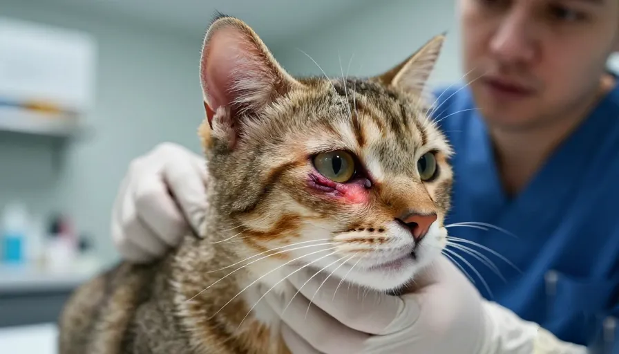
Differential Diagnostics: Dandruff vs. Pathological Dermatofitosis
A critical challenge in veterinary dermatology is distinguishing between simple, non-contagious dandruff and Dermatofitosis (Ringworm). Mistaking an active fungal infection for basic dry skin allows a highly contagious, zoonotic disease to spread unchecked through a household.
[Inspect Scaling/Alopecia] ──► Execute Wood's Lamp Examination (365 nm)
                                             │
                                             ▼
       +-------------------------------------+-------------------------------------+
       |                                                                           |
 [ Positive Apple-Green Fluorescence ]                                       [ Negative Fluorescence ]
 - Confirms Pteridine metabolite synthesis.                                  - Collect epidermal scales.
 - Indication of active Microsporum canis.                                   - Perform DTM Fungal Culture.
 - Initiate strict fungal isolation protocol.                                - Evaluate for basic Dandruff/Parasites.
3.1 Pathophysiology of Dermatofitosis (Microsporum canis)
Dermatofitosis is an infectious superficial fungal infection caused primarily by Microsporum canis in felines. Unlike dandruff, which is an internal breakdown of the skin cycle, dermatophytes are specialized parasites that consume keratin.
The fungal spores (arthroconidia) attach to the skin surface, germinate, and invade the hair shaft and stratum corneum. As the hyphae tunnel through the hair follicle, they structurally weaken the keratin matrix, causing the hair to break off at the base. This results in the classic clinical presentation: focal, expanding patches of hair loss (alopecia) covered in thick, silver-white crusts and scales.
3.2 Advanced Diagnostic Protocols for Clinical Differentiation
To avoid diagnostic errors, veterinarians rely on a structured testing protocol:
-
Wood’s Lamp Luminescence: The clinician exposes the affected coat to a specialized ultraviolet light source filtered through a Wood’s filter ($\approx 365\text{ nm}$ wavelength). In roughly 50% to 80% of Microsporum canis infections, the fungus produces a specific chemical metabolite called pteridine. Under the lamp, this metabolite emits a bright, unmistakable apple-green fluorescence along the infected hair shafts. Benign dandruff, by contrast, will either show no reaction or emit a dull, chalky white glow from soap residues or lint.
====================================================================
                  [ THE DIAGNOSTIC MATRIX ]
====================================================================
  Clinical Vector      Benign Feline Dandruff    Dermatofitosis (Ringworm)
--------------------------------------------------------------------
  Primary Cause        EFA deficit, low humidity  Infectious Keratinophilic Fungus
  Hair Involvement     Intact hair shafts        Broken, fragile hair shafts
  Lesion Pattern       Diffuse dorsal scaling    Circular focal alopecia
  Zoonotic Hazard      Zero Risk                 Extremely High Contagion
  DTM Media Color      No color transition       Rapid Amber-to-Red shift
====================================================================
-
Dermatophyte Test Medium (DTM) Culture: The definitive test remains a fungal culture using specialized Dermatophyte Test Medium. The clinician plucks suspect hairs and scrapes scaling debris into a vial containing a nutrient agar enhanced with phenol red pH indicators. Because dermatophytes preferentially metabolize alkaline proteins first, an active infection will turn the agar from amber to bright red within 3 to 14 days, concurrently with visible colony growth. Saprophytic fungi (which can contaminate regular dandruff) consume carbohydrates first and will only trigger a color change after massive colony growth, allowing for clear clinical differentiation.
The Pathophysiology of “Walking Dandruff” (Cheyletiellosis)
When evaluating a scaling cat, the clinician must always rule out an active external parasite infestation. One of the most common parasitic culprits is Cheyletiellosis, an infestation caused by the obligate mite Cheyletiella blakei.
[Cheyletiella Mite Activity] ──► [Sub-Epidermal Tunnelling / Feeding]
                                            │
                                            ▼
[Massive Exfoliative Scaling] ──► [Mites Transport Scales Along Dorsum]
                                            │
                                            ▼
[Visual Sign: "Walking Dandruff" Phenomenon] ──► [Severe Pruritus & Excoriation]
4.1 Morphological Characteristics and Behavior of Cheyletiella Mites
Cheyletiella mites are large, non-burrowing parasites that live entirely within the superficial keratin layer of the skin. Morphologically, they are distinguished by prominent, curved, hook-like accessory palpal claws near their mouthparts, which they use to anchor themselves to the epidermis while feeding on tissue fluids and surface debris.
As these mites move through the keratin layers, their feeding activity causes intense local irritation and a dramatic spike in skin cell production, resulting in a large volume of loose, white scales. The mites frequently crawl beneath these large sheets of dander, transporting them across the hair coat. This creates the unique visual phenomenon that gives the parasite its common name: “walking dandruff.”
4.2 Clinical Impact and Diagnostic Tracking
Unlike simple, non-pruritic dandruff caused by dry air, Cheyletiellosis triggers intense itching (pruritus). Affected cats will scratch, bite, and over-groom obsessively, often leading to raw, broken skin (excoriations), secondary bacterial infections, and patches of self-induced hair loss along the back.
====================================================================
              [ DIAGNOSTIC TRACE FOR CHEYLETIELLOSIS ]
====================================================================
  [Step 1]  == Clear Acetate Tape Impression applied to scaling coat.
               │
  [Step 2]  == Secure tape sample flat onto a clear glass slide.
               │
  [Step 3]  == Examine under low-power Microscopic Field (4x - 10x).
               │
  [Target]  == Detect large mites with distinct curved palpal hooks.
====================================================================
Diagnosing the condition requires a simple acetate tape impression. The clinician presses clear adhesive tape firmly against the scaling areas to trap the parasites and debris. The tape is then mounted flat onto a glass slide and examined under a microscope at low power ($4\times\text{ to }10\times$). Finding the distinctive mites or their oval eggs attached to hair shafts confirms the diagnosis, ruling out simple dry skin and guiding the appropriate antiparasitic treatment.
Structural Nutritional Architecture and Treatment Protocols
Once the diagnostic process has ruled out fungal and parasitic infections, resolving idiopathic feline dandruff requires a multi-layered approach targeting nutrition, direct skin care, and environmental management.
[Diagnose Feline Dandruff] ──► Optimize EFA Ratios (High EPA/DHA Marine Lipids)
                                             │
                                             ▼
     +---------------------------------------+---------------------------------------+
     |                                                                               |
 [ Targeted Topical Hydration ]                                      [ Macro-Volumetric Fluid Intake ]
 - Apply humectant Spot-On therapies.                                - Transition dry diet to premium Wet Food.
 - Comb 2-3x weekly to distribute sebum.                            - Install multi-fountain hydration points.
 - Utilize specific oatmeal-based rinse.                             - Restore optimal intracellular turgor.
5.1 Rebalancing Essential Fatty Acids with Marine Lipids
To rebuild a damaged skin barrier from the inside out, the diet must be enriched with highly bioavailable polyunsaturated fats. While plant oils like flaxseed contain Omega-3s in the form of Alpha-Linolenic Acid (ALA), cats lack the metabolic pathways to efficiently convert ALA into active, anti-inflammatory compounds.
The diet must instead include direct sources of animal-derived marine lipids:
-
Eicosapentaenoic Acid (EPA): Competes with arachidonic acid at the cellular level, safely blocking inflammatory pathways and reducing skin redness and irritation.
-
Docosahexaenoic Acid (DHA): Integrates directly into the phospholipid membranes of epidermal cells, strengthening the structural integrity of the stratum corneum and cutting down trans-epidermal water loss.
These target lipids are found in high concentrations within premium wild-caught cold-water fish oils (such as salmon, anchovy, and sardine oils). Supplementing the diet with these oils effectively restores the skin’s natural “mortar” layer, resolving dry flaking at its source.
5.2 Hydration Strategies: The Wet Food Advantage
Chronic low-grade dehydration is a common, overlooked driver of flaky skin. Because domestic cats evolved from desert-dwelling wild ancestors, they possess a naturally weak internal thirst drive, relying on their prey to meet their fluid needs.
When fed an exclusive diet of dry kibble ($\approx 10\%$ moisture content), cats often live in a state of mild, chronic dehydration, which shows up outwardly as dry, inelastic skin and dander.
Transitioning the cat to high-quality, protein-dense wet food formulations ($\ge 75\%$ moisture content) increases their daily water intake directly with their meals. This systemic hydration restores optimal moisture balance to the skin cells, supporting a healthy, normal keratinization cycle.
5.3 Topical Care and Environmental Controls
-
Targeted Topical Spot-Ons: For immediate relief, apply specialized dermatological spot-on pipettes containing a concentrated blend of essential fatty acids, ceramides, and cholesterol directly to the skin. These topicals spread naturally across the lipid layer, instantly patching holes in the skin barrier while systemic nutritional changes take effect.
-
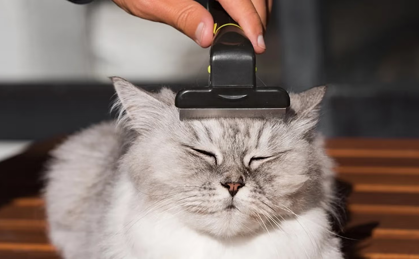
Grooming Support: Assist cats with mobility issues by combing them two to three times a week with a fine-toothed slicker brush. This manual care mimics natural grooming, sweeping away dead hair and dander while gently stimulating the sebaceous glands to distribute healthy oils along the coat.
-
Humidity Control: Use an ultrasonic, cool-mist humidifier in the rooms where your cat spends the most time. Keeping ambient humidity levels consistently between 50% and 60% prevents the surrounding air from drying out the skin, providing long-term protection against dry weather dander.
Holistic Companion Animal Health and Multi-Species Preventative Protocols
A true, comprehensive approach to pet wellness recognizes that managing dermatological issues is just one aspect of a larger care framework. Protecting our companion animals requires balancing skin care, behavioral management, specialized life-stage nutrition, and preventative veterinary medicine across all species in the home.
====================================================================
                  [ THE SYSTEMIC WELLNESS SPECTRUM ]
====================================================================
  [Feline Focus]   ──► Manage Dermatological Barriers & Stress Cytokines.
  [Canine Focus]   ──► Maintain Structural Monocoat Hygenic Routines.
  [Surgical Focus] ──► Caloric Regulation Post-Metabolic Gonadectomy.
====================================================================
6.1 Feline Lower Urinary Tract Disease (FLUTD) and Stress Management
Just as poor nutrition can cause skin flaking, environmental stress can trigger physical illness inside the cat. A prime example is Feline Lower Urinary Tract Disease (FLUTD), which includes conditions like Feline Idiopathic Cystitis (FIC).
When a cat experiences ongoing stress—such as territorial tension in a multi-pet home, sudden changes in the household, or competitive feeding setups—their central nervous system triggers a release of inflammatory chemicals. These chemicals damage the protective glycosaminoglycan (GAG) lining of the inner bladder wall.
Once this layer is compromised, highly concentrated urine directly irritates the sensitive underlying tissue, causing painful inflammation, bloody urine (hematuria), straining in the litter box, and life-threatening urethral blockages in male cats.
[Chronic Environmental Stress] ──► Bladder GAG Layer Degradation ──► Local Sterile Cystitis (FLUTD)
Preventing these painful urinary crises requires a dedicated strategy to reduce stress and boost hydration:
-
Continuous Fluid Access: Place multiple running water fountains around the home to encourage drinking and dilute the urine.
-
Targeted Urinary Diets: Feed specialized urinary formulas (such as Pro Plan Urinary Care). These diets are carefully engineered to restrict magnesium and phosphorus levels while keeping urine pH in the ideal 6.0 to 6.3 range, preventing the formation of painful struvite and calcium oxalate stones.
6.2 Advanced Canine Husbandry: The Poodle Matrix
A comprehensive approach to pet care also means adapting to the unique biological needs of different breeds. For example, the Poodle (Canis lupus familiaris) possesses a highly distinct anatomical and physical matrix that demands specialized routine care.
                       [ THE CANINE COAT AND ORAL AXIS ]
                                       │
       +-------------------------------+-------------------------------+
       |                                                               |
 [ Non-Shedding Monocoat Dynamics ]                          [ Periodontal Defense Protocol ]
 - Continuous follicular growth phase.                       - Narrow mandibular structure crowds teeth.
 - Traps dead hair, causing severe matting.                  - High plaque calculus accumulation rate.
 - Demands daily systematic slicker brushing.                - Requires home brushing & annual scaling.
-
Dermatological and Coat Management: Poodles have a single-layered monocoat that grows continuously without shedding. While this makes them ideal for households with allergies, it presents a significant maintenance challenge. Because dead hairs cannot fall out naturally, they become trapped within the tight curls, rapidly forming dense, painful mats that pull against the skin and trap moisture. Left unchecked, these mats can lead to severe bacterial hot spots and yeast dermatitis. Managing a Poodle requires daily brushing down to the skin with a high-quality slicker brush, combined with professional clipping and grooming every 4 to 6 weeks.
-
The Auditory Canal Matrix: Poodles also grow dense hair deep inside their external ear canals. This hair traps wax, debris, and moisture, creating an ideal environment for painful yeast and bacterial ear infections (otitis externa). Owners must clean the ears weekly using a dedicated drying solution and gently pluck or trim excess internal hair to ensure proper airflow.
-
Preventative Dental Prophylaxis: Due to their narrow, elegant snout structure, Miniature and Toy Poodles have crowded teeth, creating perfect pockets that trap food and bacteria. They accumulate plaque and tartar at a significantly faster rate than larger breeds. To prevent advanced periodontal disease, owners should brush their dog’s teeth at least three times a week with enzymatic pet toothpaste and schedule annual professional veterinary cleanings under anesthesia. This preventative care is critical; unchecked oral bacteria can eventually enter the bloodstream, causing permanent, life-threatening damage to the heart valves, liver, and kidneys.
-
Tailored Phase Nutrition: Support your Poodle’s long-term health by providing high-quality, breed-appropriate nutrition, such as PRO PLAN Small & Toy Puppy with bovine colostrum to strengthen their immune defenses, transitioning later to PRO PLAN Small & Toy Adult to fuel their fast metabolism and maintain lean, strong muscles.
6.3 The Pathophysiology and Aftercare of Surgical Sterilization
Surgical sterilization—ovariohysterectomy for females and orchiectomy for males—is a vital, responsible procedure that prevents unwanted litters, eliminates reproductive cancers, and stops behaviors like territorial roaming and urine spraying. However, removing the gonads causes a massive hormonal shift that directly impacts the animal’s metabolism.
[Surgical Gonadectomy] ──► Depletion of Estrogen / Testosterone ──► 20-30% Drop in BMR
                                                                            │
                                                                            ▼
[Portions Unchanged] ──► Pathological Adipose Deposition ──► Chronic Insulin Resistance (Diabetes)
Within days of surgery, the loss of circulating estrogen or testosterone causes the animal’s Basal Metabolic Rate (BMR) to drop by 20% to 30%. At the same time, this hormonal change alters the brain’s satiety signals, making the pet feel hungrier. If owners do not carefully manage their pet’s food, this combination leads to rapid weight gain and obesity.
Excess fat tissue acts as an active endocrine organ, secreting a constant stream of inflammatory signals (adipokines) that down-regulate insulin receptors, frequently leading to Type-2 Feline Diabetes Mellitus.
To protect a sterilized pet’s health, implement a strict care and feeding plan:
-
Immediate Caloric Regulation: Reduce daily food portions by 20% to 30% right after surgery, or transition the pet to a specialized Sterilized/Weight Management diet that features a high-fiber, low-calorie profile to keep them feeling full without adding excess weight.
-
Strict Portion Weighing: Stop free-feeding. Use a digital kitchen scale to weigh out precise portions to the gram, adjusting the amount based on regular weight checks.
-
Post-Operative Recovery Care: During the critical 10-to-14-day post-op healing window, enforce strict crate rest and leash-only walks to prevent running or jumping from tearing internal sutures. Perform daily checks of the incision site for any redness, heat, or discharge, and ensure the pet wears an Elizabethan collar (E-collar) continuously to prevent licking, which can introduce mouth bacteria and cause serious infection.
Comprehensive Comparative Reference Matrix
To assist owners and veterinary professionals in making quick, accurate clinical decisions, the following matrix outlines the diagnostic profiles, primary causes, and immediate intervention strategies for common pet health and wellness challenges.
| Clinical Condition | Primary Visual / Diagnostic Indicators | Core Underlying Driver | Immediate Remedial Protocol | Long-Term Preventative Strategy |
| Benign Feline Dandruff | Diffuse white flakes along the dorsal line; hair shafts remain strong and intact; zero itching or skin redness. | Deficiencies in essential fatty acids (Omega-3/6); chronically dry indoor air; or reduced grooming due to obesity or arthritis. | Increase dietary intake of high-quality marine lipids (EPA/DHA) and incorporate wet food formulas. | Install a cool-mist humidifier to maintain 50-60% humidity; brush the coat 2-3 times weekly; manage weight. |
| Dermatofitosis (Ringworm) | Focal, expanding patches of hair loss (alopecia); thick silver crusts; bright apple-green fluorescence under a Wood’s Lamp. | Superficial fungal infection (Microsporum canis) that consumes keratin within the hair shafts and outer skin layer. | Isolate the animal immediately to protect others; consult a vet for targeted topical and oral antifungal treatments. | Perform deep environmental disinfection to clear fungal spores; run a DTM culture to confirm complete recovery. |
| Cheyletiellosis (Walking Dandruff) | Massive scaling along the back; flakes appear to move; accompanied by intense itching, scratching, and raw skin. | Infestation by the surface-dwelling mite Cheyletiella blakei, which feeds on tissue fluids and disrupts the skin layer. | Apply a vet-approved, species-specific topical antiparasitic treatment (e.g., Selamectin or Fipronil). | Treat all contact animals in the home simultaneously; thoroughly wash and sanitize all bedding and carpets. |
| Urinary Distress (FLUTD / FIC) | Frequent, unproductive trips to the litter box; bloody urine (hematuria); crying or straining while attempting to urinate. | Stress-induced breakdown of the protective glycosaminoglycan (GAG) layer lining the inner bladder wall. | Emergency veterinary care for any male cat to clear or prevent life-threatening urethral blockages. | Transition to high-moisture wet foods; keep urine pH stable at 6.0–6.3; use pheromone diffusers to lower stress. |
| Canine Monocoat Matting (Poodles) | Tight, dense tangles of hair forming close to the skin surface; localized redness; frequent head shaking. | Continuous hair growth without natural shedding, causing dead hairs to become permanently trapped in the curls. | Carefully split and tease out mats using a slicker brush and detangling spray; treat skin hot spots with antiseptics. | Commit to daily thorough brushing down to the skin; schedule professional clips and ear cleanings every 4-6 weeks. |
| Post-Sterilization Metabolic Shift | Rapid accumulation of body fat along the abdomen; increased food seeking; low energy or decreased activity. | A sudden 20% to 30% drop in Basal Metabolic Rate (BMR) caused by the removal of reproductive hormones. | Instantly reduce daily calorie portions by 20% to 30% or switch to a specialized weight management diet. | Stop free-feeding; weigh portions to the gram using a digital scale; schedule 30 minutes of active daily pl |
FAQ – Feline Abscesses: Causes, Symptoms, Treatment, and Prevention
1. What is a feline abscess?
A feline abscess is a localized pocket of pus that develops beneath the skin or within tissues after bacteria enter the body through a wound, bite, scratch, or dental infection. The body’s immune system attempts to isolate the infection by creating a capsule around the affected area, leading to the formation of a swollen, painful lump filled with purulent material.
2. What causes abscesses in cats?
The most common causes include:
- Bite wounds from cat fights.
- Deep scratches from other animals.
- Puncture wounds from sharp objects.
- Dental infections affecting tooth roots.
- Contaminated skin injuries.
- Foreign objects trapped beneath the skin.
Common bacteria involved include Pasteurella multocida, Bacteroides species, Fusobacterium species, and Streptococcus species.
3. Why do cat bite wounds often become abscesses?
Cat teeth are long, narrow, and sharp, creating deep puncture wounds that quickly seal at the skin surface. This traps bacteria beneath the skin, creating a warm, oxygen-poor environment where bacteria multiply rapidly, eventually producing an abscess.
4. What are the first signs of an abscess in cats?
Early signs may include:
- Small swelling beneath the skin.
- Pain when touched.
- Redness around the wound.
- Warm skin over the affected area.
- Mild lethargy.
- Reduced appetite.
- Sensitivity during movement.
These signs often appear several days after the initial injury.
5. What does an abscess feel like?
An abscess usually feels:
- Soft or fluid-filled.
- Warm to the touch.
- Painful when pressed.
- Round or irregularly shaped.
- Movable beneath the skin.
Veterinarians often describe this as a “fluctuant swelling.”
6. Can an abscess burst on its own?
Yes. As pressure builds inside the abscess, the skin may rupture and release:
- Thick yellow pus.
- Greenish discharge.
- Blood-stained fluid.
- Foul-smelling material.
Although drainage may temporarily relieve pressure, veterinary treatment is still necessary to eliminate the infection completely.
7. Where do abscesses most commonly occur on cats?
Common locations include:
- Face and cheeks.
- Neck.
- Tail base.
- Front legs.
- Hind legs.
- Back and shoulders.
These areas are frequently targeted during territorial fights.
8. What is a dental abscess in cats?
A dental abscess develops when bacteria invade the tooth root and surrounding bone structures. It often causes:
- Facial swelling.
- Bad breath.
- Excessive drooling.
- Difficulty eating.
- Pawing at the mouth.
- Swelling beneath the eye.
Dental abscesses require veterinary treatment and often tooth extraction.
9. Can a cat abscess be dangerous?
Yes. Without treatment, bacteria may spread into the bloodstream and cause:
- Sepsis.
- Tissue necrosis.
- Osteomyelitis (bone infection).
- Chronic pain.
- Systemic illness.
- Organ complications.
Prompt treatment significantly reduces these risks.
10. Should I squeeze or drain an abscess at home?
No. Attempting to squeeze or puncture an abscess can:
- Cause severe pain.
- Spread bacteria deeper into tissues.
- Increase inflammation.
- Delay healing.
- Introduce new infections.
Abscess drainage should only be performed under veterinary supervision.
11. How do veterinarians treat feline abscesses?
Treatment generally includes:
- Sedation or anesthesia.
- Surgical opening of the abscess.
- Complete drainage of pus.
- Removal of dead tissue.
- Sterile flushing of the cavity.
- Placement of a drain when necessary.
- Antibiotic therapy.
- Pain management medications.
12. What is a Penrose drain?
A Penrose drain is a soft latex tube inserted into deep abscess cavities to:
- Allow continuous drainage.
- Prevent premature skin closure.
- Reduce fluid accumulation.
- Improve healing outcomes.
Drains are usually removed after 3 to 5 days.
13. How long does it take for an abscess to heal?
Recovery times vary:
- Small abscesses: 7–14 days.
- Moderate abscesses: 2–3 weeks.
- Extensive abscesses: Several weeks.
Proper wound care and medication compliance significantly improve healing speed.
14. Will my cat need antibiotics?
Most cats require antibiotics to eliminate remaining bacteria after drainage. Commonly prescribed medications include:
- Amoxicillin-Clavulanate.
- Cefovecin sodium.
- Other veterinarian-selected broad-spectrum antibiotics.
Always complete the full course of treatment.
15. Can indoor cats develop abscesses?
Yes. Although outdoor cats are at higher risk, indoor cats may develop abscesses from:
- Household accidents.
- Dental disease.
- Scratches from other pets.
- Embedded foreign objects.
- Self-inflicted wounds.
16. Why do veterinarians recommend FIV and FeLV testing after cat fights?
Cat bites are a major transmission route for:
- Feline Immunodeficiency Virus (FIV).
- Feline Leukemia Virus (FeLV).
Testing approximately 60 days after a fight helps determine whether viral transmission occurred.
17. How can I prevent abscesses in my cat?
Preventive measures include:
- Keeping cats indoors.
- Supervising outdoor access.
- Using secure cat enclosures.
- Sterilizing cats to reduce fighting behavior.
- Maintaining vaccinations.
- Promptly treating wounds.
- Scheduling routine veterinary exams.
18. Does neutering reduce abscess risk?
Yes. Neutering and spaying significantly reduce:
- Territorial aggression.
- Roaming behavior.
- Fighting frequency.
- Bite-related injuries.
Studies and veterinary experience indicate a substantial reduction in abscess incidence after sterilization.
19. What should I feed a cat recovering from an abscess?
Recovery nutrition should emphasize:
- High-quality animal protein.
- Adequate hydration.
- Essential fatty acids.
- Vitamins and minerals that support tissue repair.
- Easily digestible ingredients.
Life-stage appropriate diets are important for kittens, adults, and senior cats.
20. When is an abscess considered an emergency?
Seek immediate veterinary care if your cat experiences:
- High fever.
- Severe facial swelling.
- Difficulty breathing.
- Inability to eat or drink.
- Extreme lethargy.
- Persistent bleeding.
- Rapidly expanding swelling.
- Signs of sepsis or collapse.
 (1)")
