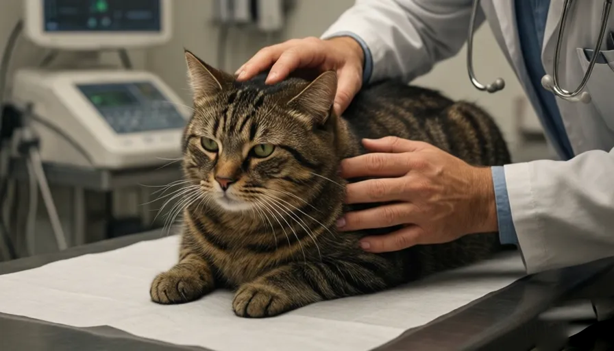
Hearing your veterinarian recommend a biopsy for your beloved cat can send a sudden spike of anxiety through any pet parent’s heart. Your mind might immediately jump to worst-case scenarios, painting scary pictures of complex invasive surgeries, heavy general anesthesia, and painful recoveries.
However, a biopsy is one of the most powerful, precise, and compassionate diagnostic tools available in modern veterinary medicine. Rather than being a reason to panic, a biopsy is a definitive roadmap. It takes the guesswork out of your cat’s symptoms, allowing your veterinary team to move past assumptions and prescribe the exact treatment plan your cat needs.
In this comprehensive guide, we will unpack everything you need to know about feline biopsies. We’ll break down the structural differences between cellular and tissue samples, detail the various collection methods used by vets, map out what to expect during recovery, and provide a realistic overview of financial costs. Our goal is to transform this clinical unknown into a manageable, transparent step toward healing your companion.
Demystifying the Biopsy Cells vs. Architecture
To truly understand what a biopsy is, we must look at how veterinarians analyze disease at a microscopic level. In the broadest medical terms, a biopsy is the collection of a sample of cells or tissues from a living body to be evaluated by a diagnostic specialist.
When your cat develops a mysterious lump, chronic skin irritation, or an enlarged internal organ, a veterinarian cannot determine the cause simply by looking at or feeling the surface. They must look at the microscopic blueprints. Depending on how the sample is harvested, veterinary diagnostics are split into two major categories: Cytology and Histopathology.
                  THE MICROSCOPIC DIAGNOSTIC SPLIT

  [ Cytology ]       ──► Examines loose, isolated cells (Fried egg view)

  [ Histopathology ] ──► Examines intact structural tissue (Three-dimensional blueprint)
1. Cytology: The Two-Dimensional Cellular Snapshot
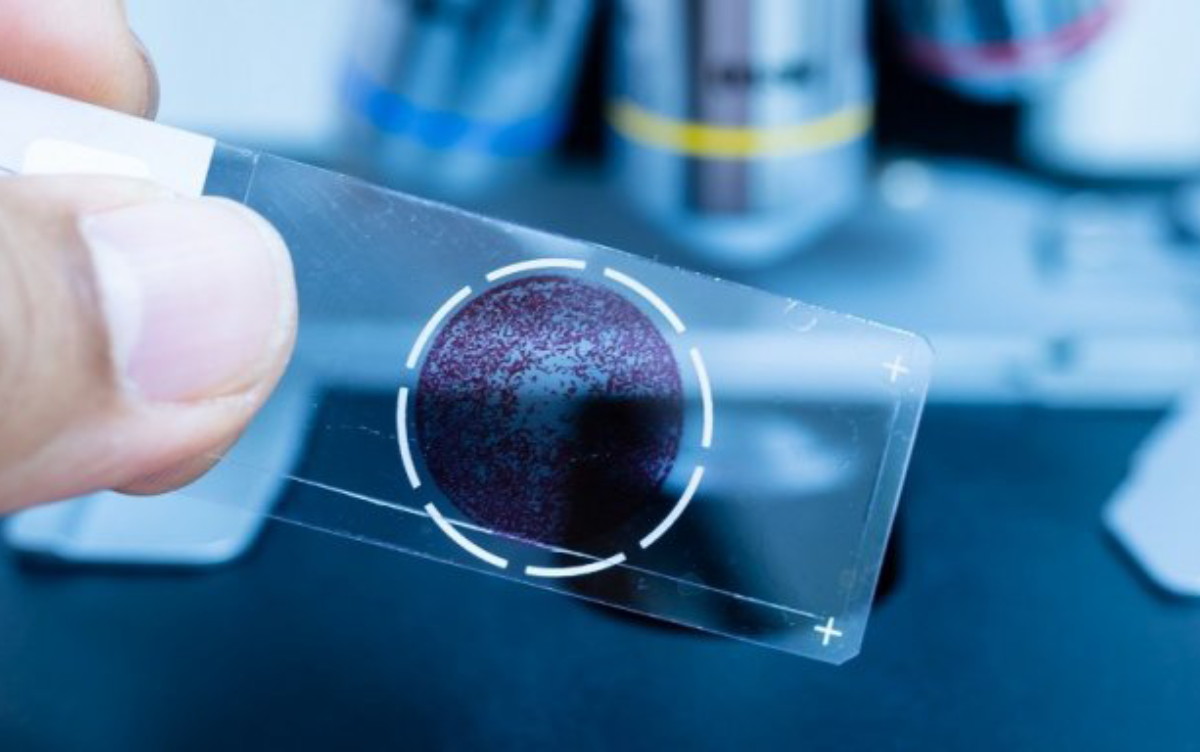
When a veterinarian collects loose cells from a mass using a thin needle and a syringe, the process is called a Fine Needle Aspirate (FNA). The resulting laboratory evaluation is known as cytology.
Think of cytology as a two-dimensional view of individual cells spread out across a flat glass slide. Pathologists describe this as looking at a “fried egg.” You can clearly see the nucleus (the egg yolk) and the surrounding cytoplasm (the egg white), but all the cells are separated from each other.
-
The Advantage: It is minimally invasive, highly cost-effective, and usually performed during a standard exam without any sedation.
-
The Limitation: Because the cells are loose and detached, a pathologist cannot see how they interact with neighboring tissues. This lack of structural context can sometimes lead to inconclusive or incomplete results.
2. Histopathology: The Gold Standard Tissue Architecture
True tissue histopathology is the primary focus of a formal biopsy. Instead of harvesting a few loose cells, the veterinarian surgically removes an intact piece of solid tissue or an entire mass.
This sample is placed in a chemical preservative solution called formalin to halt biological decay (a process called fixation). A laboratory technician then embeds the hardened tissue in blocks of paraffin wax, slices it into microscopic sections, and stains it with specialized dyes.
Pathologists view this as a three-dimensional examination. Rather than looking at a flat “fried egg,” they are evaluating a cross-section of a “hard-boiled egg.”
-
The Advantage: Histopathology preserves the structural architecture of the tissue. It allows a board-certified veterinary pathologist to see exactly how cells are arranged, whether they are invading nearby blood vessels, and how the surrounding healthy tissue is reacting. This structural context provides a definitive, highly accurate diagnosis.
The Core Types of Feline Tissue Biopsies
Veterinarians choose specific biopsy collection methods based on the size, location, and suspected severity of the tissue anomaly. These techniques are categorized by how much tissue is harvested and the specialized tools required.
                     FELINE TISSUE BIOPSY TAXONOMY

  [ Punch Biopsy ]     ──► Circular blade ──► Best for skin and surface lesions.
  [ Incisional ]       ──► Partial wedge  ──► Best for large masses or internal organs.
  [ Excisional ]       ──► Total removal  ──► Best for treating while diagnosing.
  [ Needle/Tru-Cut ]   ──► Narrow core    ──► Best for internal ultrasound-guided targets.
1. Punch Biopsy
A punch biopsy utilizes a specialized medical instrument featuring a sharp, circular, pen-like blade. The veterinarian presses the tool downward into the affected area and rotates it slightly to core out a small, cylindrical plug of skin.
-
Primary Application: Diagnosing unusual skin conditions, non-healing sores, autoimmune skin disorders, or very small surface growths.
-
Dimensional Scope: These samples are small, typically ranging from 4 to 8 millimeters in diameter, and usually require just one or two small sutures to close.
2. Incisional (Wedge) Biopsy
An incisional biopsy involves surgically carving out a small, wedge-shaped piece of tissue from a much larger mass or internal organ using a scalpel or surgical scissors.
-
Primary Application: Screening large, deeply embedded tumors or evaluating internal organs like the liver, kidneys, or spleen.
-
Strategic Purpose: The goal is to safely discover exactly what a disease process is before committing to a massive, highly invasive surgery or intensive chemotherapy.
3. Excisional Biopsy
An excisional biopsy combines diagnostic evaluation with active treatment. During this surgical procedure, the veterinarian removes the entire mass or suspicious growth along with a wide border of surrounding healthy tissue.
-
Primary Application: Treating distinct skin tumors, subcutaneous lumps, or affected lymph nodes.
-
Strategic Purpose: The goal is to completely cure the local condition by removing the problem area entirely, while simultaneously sending the tissue to a lab to confirm what it was and ensure no dangerous cells were left behind.
4. Needle Core Biopsies (e.g., Tru-Cut)
A needle core biopsy uses a long, specialized instrument (such as a Tru-Cut needle) to capture a thin, structural cylinder of tissue from inside the body. This is distinct from a fine needle aspirate, which only sucks up loose cells.
-
Primary Application: Harvesting samples from deep internal organs inside the abdomen or chest cavity.
-
Strategic Purpose: This technique is frequently performed with the guidance of an advanced ultrasound machine, allowing veterinarians to sample deep internal tissues safely without needing an open surgical incision.
Navigating the Three Levels of Procedural Anesthesia
Because collecting a tissue biopsy requires making an incision into real tissue, ensuring your cat feels absolutely no pain or distress is a primary medical requirement. Depending on your cat’s temperament, health status, and the location of the biopsy, your veterinarian will utilize one of three chemical management methods.
1. Local Anesthetic (The Surface Block)
An injectable numbing medication (such as lidocaine or bupivacaine) is carefully infiltrated into the skin immediately surrounding the biopsy site, completely blocking local pain signals.
-
When It’s Used: Very small, superficial skin punch biopsies or removals of tiny surface growths less than a centimeter in diameter.
-
The Catch: The cat must be extraordinarily calm, cooperative, and able to remain perfectly still while being gently held by veterinary assistants. If a cat is anxious or combative, this method is unsafe.
2. Sedation (The Relaxed Twilight State)
Sedation involves injecting a combination of fast-acting medications that make the cat deeply drowsy, relaxed, and less reactive to their surroundings, though they are not fully asleep. This is almost always paired with a local anesthetic block at the biopsy site.
-
When It’s Used: Moderately invasive skin punches, needle core biopsies, or when handling an anxious cat who would become overly stressed by simple physical restraint.
-
The Catch: While safer and less intensive than full general anesthesia, sedation still requires careful monitoring of heart rate and respiration. It is generally restricted to surface procedures.
3. General Anesthesia (Complete Controlled Sleep)
General anesthesia places your cat into a deep, completely unconscious state where they feel zero pain and have no awareness of the procedure. It requires placing an endotracheal breathing tube and utilizing advanced electronic systems to continuously monitor vital signs.
-
When It’s Used: Deep abdominal or chest exploratory surgeries, large excisional mass removals, oral biopsies inside the mouth, and specialized internal diagnostic procedures like an endoscopy.
-
The Catch: This carries the highest level of procedural complexity, requires comprehensive pre-anesthetic blood testing to ensure safety, and represents the highest financial investment.
Clinical Preparation Setting Up for Procedural Safety
Proper preparation before a biopsy is critical to minimize risks and ensure an accurate, safe procedure for your cat.
                  PRE-BIOPSY CLINICAL CHECKS

  [ Complete Blood Count ] ──► Verifies stable red/white blood cells & platelets.
  [ Chemistry Panel ]      ──► Assesses metabolic liver and kidney function.
  [ Coagulation Profile ]  ──► Mandatory safety check before internal organ sampling.
The Fasting Directives
If your cat is scheduled to undergo sedation or general anesthesia, you will receive strict instructions to withhold all food for 12 to 24 hours prior to the procedure. Fasting prevents your cat from accidentally vomiting while under the influence of anesthetic medications, removing the dangerous risk of aspiration pneumonia. Clean, fresh water is usually permitted until the morning of check-in.
Pre-Anesthetic Screening Protocols
Before administering any sedative or anesthetic agent, your vet will require a comprehensive diagnostic workup to confirm your cat’s organs can safely process the medications:
-
Complete Blood Count (CBC) & Chemistry Panel: Evaluates white blood cell counts for hidden infections, ensures red blood cell levels are stable, and checks that liver and kidney functions are strong enough to metabolize and clear the anesthetic drugs.
-
Coagulation (Clotting) Profiles: This step is absolutely mandatory if your vet is performing an internal organ biopsy (such as a liver or spleen sample). Because the liver synthesizes the proteins responsible for blood clotting, an underlying liver condition can compromise your cat’s ability to stop internal bleeding. A pre-biopsy clotting test ensures it is safe to proceed.
Post-Biopsy Home Care and Recovery
Once the biopsy is complete and your cat is discharged, their recovery depends heavily on your care and management at home.
The Immediate 24-Hour Transition
When you first bring your cat home after a sedated or anesthetic procedure, they may appear disoriented, unsteady on their feet, or unusually clingy. Alternatively, some cats may choose to isolate themselves in a dark closet or under a bed.
Keep them confined to a small, warm, quiet room (like a bathroom or spare bedroom) away from other household pets and children. Provide a low-sided litter box that doesn’t require jumping, and offer a small meal—roughly half their normal portion—as anesthesia can cause mild nausea.
Protecting the Incision Site
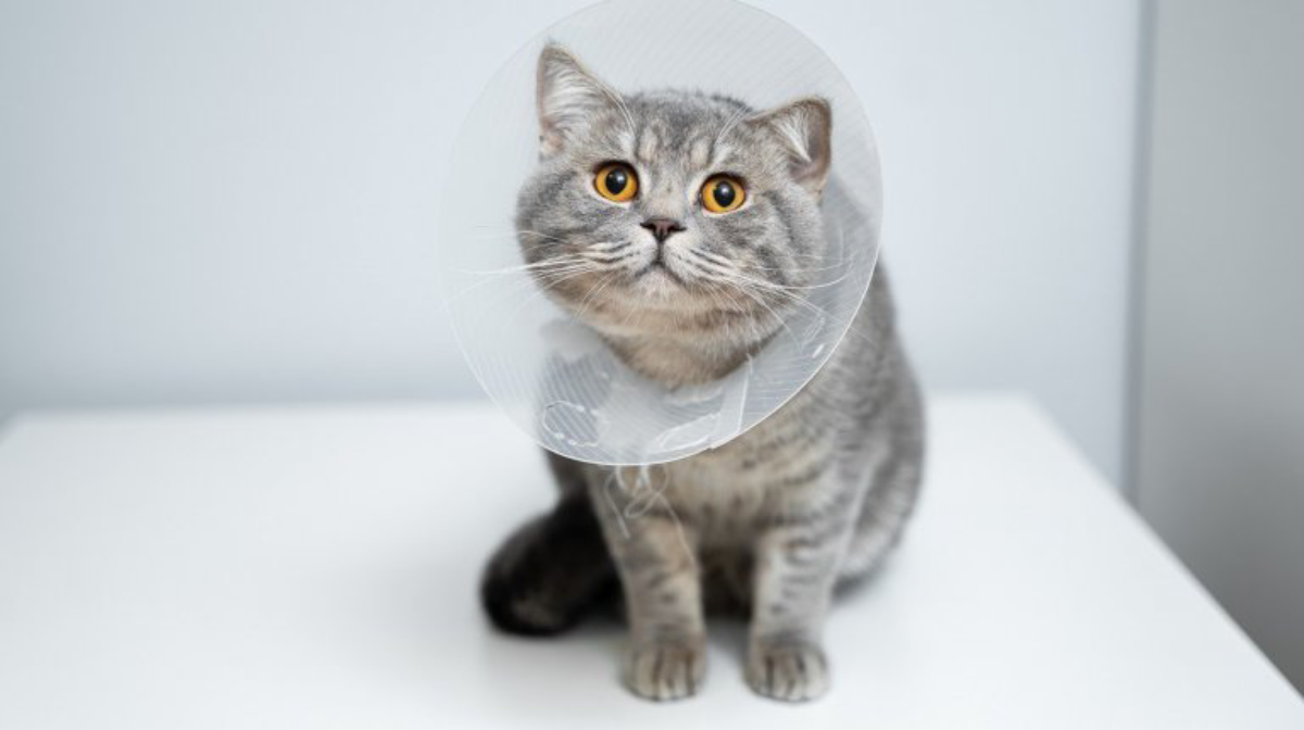
If your cat has surgical sutures or staples, you must prevent them from licking, scratching, or biting at the healing skin. A cat’s tongue is covered in backward-facing, sand-paper-like barbs that can easily rip out fresh surgical stitches or introduce bacteria from their saliva, causing a severe localized infection.
                     PROTECTIVE ARCHITECTURE EXAMPLES

  [ Elizabethan Collar ]  ──► Classic plastic cone ──► Complete physical barrier.
  [ Medical Recovery Onesie ] ──► Soft fabric suit ──► Best for abdominal incisions.
Keep your cat’s protective gear on continuously for the full 10 to 14 days of healing, or until your veterinarian clears them during their follow-up appointment.
Translating the Pathologist’s Report
Waiting for your cat’s biopsy results can be a stressful experience. While a simple fine needle aspirate cytology report might return in a few days, a formal tissue histopathology report typically takes 7 to 10 business days to process.
This delay occurs because the tissue sample must go through specialized chemical processing, wax embedding, precision slicing, and custom staining before a board-certified pathologist can evaluate it under a microscope. Bone biopsies can take even longer—often two to three weeks—because the mineralized calcium must be completely dissolved before the sample can be sliced.
When the report arrives, it is written in highly complex medical terminology. Your veterinarian will act as your translator, focusing on three critical pieces of information:
                  THE BIG THREE PATHOLOGY ANSWERS

  [ The Pathology Identity ] ──► Is it inflammatory, infectious, or neoplastic (cancer)?

  [ The Malignancy Status ]  ──► Is the tumor benign (non-spreading) or malignant (aggressive)?

  [ The Surgical Margins ]   ──► Clean Margins (Fully removed) vs. Dirty Margins (Cells remain)
Understanding Surgical Margins
If your cat underwent an excisional biopsy to remove a mass, the pathologist’s assessment of the surgical margins is incredibly important:
-
Clean (Negative) Margins: This means the pathologist found a continuous outer border of completely normal, healthy tissue surrounding the removed mass. This indicates the tumor was entirely excised, significantly reducing the chances of it growing back in that location.
-
Dirty (Positive) Margins: This means abnormal or cancerous cells were found right up to the very edge of the cut tissue. This indicates that some diseased cells likely remain inside your cat’s body, meaning further treatment—such as a second revision surgery or follow-up oncology care—may be necessary.
Financial Realities Mapping the Cost of a Feline Biopsy
The financial investment required for a feline biopsy varies widely based on the complexity of the procedure and the level of anesthesia required. Understanding these costs beforehand can help you plan and prepare.
Financial Note: These estimated cost ranges are comprehensive. They include the initial veterinary examination fee, pre-anesthetic blood testing, the surgical collection procedure, post-operative pain medications, and the laboratory fees charged by the veterinary pathologist to evaluate the tissue.
Summary Checklist for Pet Parents Facing a Biopsy
If your veterinarian has recommended a biopsy for your cat, use this step-by-step checklist to guide your conversations and preparation:
-
[ ] Clarify the Sample Type: Ask your vet if they are recommending a cellular cytology scan (Fine Needle Aspirate) or a formal tissue histopathology biopsy.
-
[ ] Understand the Anesthetic Plan: Discuss whether the sample will be harvested using a local block, twilight sedation, or full general anesthesia.
-
[ ] Complete Pre-Screening: Schedule pre-anesthetic blood work and clotting panels well ahead of the procedure date.
-
[ ] Confirm Fasting Rules: Note the exact time you must stop feeding your cat the night before the procedure.
-
[ ] Prepare a Recovery Space: Set up a quiet, low-traffic room with a low-entry litter box and fresh bedding for post-operative recovery.
-
[ ] Secure Protective Gear: Ensure you have an appropriately sized Elizabethan collar or medical recovery suit ready to protect fresh sutures.
Conclusion: A Clear Path Forward
While the word “biopsy” can sound intimidating, it is ultimately a deeply supportive tool that brings clarity to your cat’s health. It provides a bridge between noticing a symptom and providing an effective cure. By understanding the care, safety measures, and details involved in this routine veterinary procedure, you can confidently make informed decisions for your companion.
Work closely with your veterinarian, ask questions, and focus on the clear path forward that a biopsy provides. Your commitment, patience, and attentive care are the most valuable assets in helping your cat return to a happy, healthy, and comfortable life.
FAQ
1. What is a biopsy in cats?
A biopsy is a veterinary diagnostic procedure that involves collecting cells or tissue from a suspicious area of your cat’s body. The sample is examined under a microscope to determine the exact cause of a disease, lump, growth, or abnormal tissue change.
2. Why would a veterinarian recommend a biopsy?
Veterinarians recommend biopsies to diagnose unexplained lumps, tumors, chronic skin conditions, enlarged organs, persistent inflammation, mouth lesions, or other abnormalities that cannot be accurately identified through a physical exam alone.
3. Is a biopsy painful for cats?
No. Biopsies are performed using local anesthesia, sedation, or general anesthesia depending on the procedure. Your cat should not feel pain during the biopsy, and pain medications are typically provided afterward to ensure comfort.
4. What is the difference between cytology and a biopsy?
Cytology examines individual cells collected through a fine needle aspirate (FNA), while a biopsy evaluates an actual piece of tissue. Biopsies generally provide more detailed and definitive information because tissue architecture remains intact.
5. What is a fine needle aspirate (FNA)?
A fine needle aspirate uses a thin needle to collect loose cells from a lump or organ. It is minimally invasive, often performed without anesthesia, and commonly serves as the first diagnostic step before a surgical biopsy.
6. How long does a cat biopsy procedure take?
The collection procedure itself may take anywhere from 15 minutes to several hours, depending on the location and complexity of the biopsy. Most cats return home the same day unless extensive surgery is required.
7. Will my cat need anesthesia for a biopsy?
Many biopsies require sedation or general anesthesia, especially when internal organs or larger tissue samples are involved. Small skin biopsies may sometimes be performed using only a local anesthetic.
8. How should I prepare my cat for a biopsy?
Your veterinarian may recommend fasting your cat for 12 to 24 hours before the procedure. Pre-anesthetic blood tests are often performed to evaluate overall health and ensure anesthesia safety.
9. How long does it take to get biopsy results?
Most histopathology reports are available within 7 to 10 business days. Specialized biopsies, such as bone samples, may require two to three weeks due to additional processing requirements.
10. What do “benign” and “malignant” mean?
A benign growth is non-cancerous and typically does not spread to other parts of the body. A malignant growth is cancerous and may invade nearby tissues or spread to distant organs.
11. What are clean surgical margins?
Clean margins mean the pathologist found normal tissue surrounding the removed mass, suggesting the abnormal tissue was completely removed during surgery.
12. What are positive or dirty margins?
Positive margins indicate abnormal or cancerous cells were found at the edge of the removed tissue sample. This may mean some disease remains and additional treatment could be necessary.
13. How long does biopsy recovery take?
Most skin biopsy sites heal within 10 to 14 days. Recovery from larger surgical biopsies may take several weeks, depending on the tissue involved and your cat’s overall health.
14. Will my cat need to wear a cone after a biopsy?
In many cases, yes. An Elizabethan collar (cone) or recovery suit helps prevent licking, scratching, or damaging the surgical site during healing.
15. What complications can occur after a biopsy?
Complications are uncommon but may include swelling, bruising, infection, bleeding, delayed wound healing, or reactions to anesthesia. Following your veterinarian’s aftercare instructions greatly reduces these risks.
16. How much does a cat biopsy cost?
Costs vary widely depending on the procedure type, anesthesia requirements, diagnostic testing, and laboratory fees. Simple skin biopsies are generally less expensive than internal organ or surgical biopsies.
17. Can a biopsy diagnose cancer in cats?
Yes. A biopsy is considered one of the most reliable methods for diagnosing cancer, determining tumor type, evaluating aggressiveness, and helping veterinarians develop an appropriate treatment plan.
18. Is a biopsy always necessary if my cat has a lump?
Not always. Veterinarians often start with a fine needle aspirate. If the results are inconclusive or more detailed information is needed, a biopsy may then be recommended.
19. Can older cats safely undergo a biopsy?
Many senior cats safely undergo biopsies every year. Pre-anesthetic testing helps veterinarians assess risks and customize anesthesia protocols based on your cat’s age and medical condition.
20. What happens after the biopsy results come back?
Your veterinarian will review the pathology report, explain the diagnosis, discuss treatment options, and create a personalized care plan based on the findings.