The Comprehensive Guide to Feline Neoplasia: Clinical Pathology, Diagnostic Frameworks, and Multimodal Therapeutics
2 weeks ago
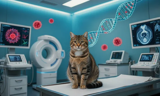
Neoplasia in felines represents a major challenge in modern veterinary oncology. Statistically, approximately one in five cats will face a neoplastic diagnosis during their lifetime. As advancements in veterinary nutrition, indoor husbandry, and preventative medicine extend the average feline lifespan, the incidence of age-related cancers has risen. In felines over ten years of age, neoplasia is a leading cause of mortality.
For veterinary clinicians, technicians, and dedicated owners, identifying neoplastic disease requires a solid understanding of oncology. Unlike some companion animals where benign lipomas are common, cellular growths in cats are frequently highly invasive and malignant.
This guide explores the pathophysiology of common feline tumors, analyzes environmental and viral triggers, details diagnostic staging protocols, and breaks down modern multimodal treatment strategies designed to prolong quality of life.
Understanding Feline Neoplasia at the Cellular Level
At its core, neoplasia is an abnormal proliferation of cells that escapes the body's regulatory mechanisms. In healthy tissue, cellular reproduction is tightly governed by a balance between oncogenes (which promote cell growth) and tumor suppressor genes (which trigger apoptosis, or programmed cell death, when DNA damage is detected).
               [The Transformation to Cellular Malignancy]
                                   │
         ┌─────────────────────────┴─────────────────────────┐
         ▼                                                   ▼
   [Benign Tumors]                                   [Malignant Tumors]
• Localized growth capsule                         • Infiltrative, unencapsulated
• Does not invade adjacent tissues                 • Destroys local tissue architecture
• Rare metastasis potential                        • High risk of vascular/lymphatic spread
When genetic mutations disrupt this balance, cells divide uncontrollably, leading to tumor formation. These growths are classified into two main categories:
Benign Neoplasms
Benign tumors grow locally and are often contained within a distinct tissue capsule. They do not invade surrounding tissues or spread (metastasize) to distant organs. While they are typically not life-threatening, benign tumors can still cause clinical issues if their physical size presses against vital structures like brain tissue, blood vessels, or the urinary tract.
Malignant Neoplasms (Cancer)
Malignant tumors are unencapsulated, aggressive, and highly invasive. They lack clear boundaries, sending microscopic strands of cancer cells directly into surrounding healthy tissue.
As these tumors grow, they break through local tissue barriers and enter the blood vessels or lymphatic system. From there, they travel to distant organs—such as the lungs, liver, or regional lymph nodes—and establish secondary tumor sites (metastasis).
Major Pathological Classifications of Feline Tumors
Feline oncology differs significantly from canine oncology. Cats have a lower overall incidence of skin tumors than dogs, but when a tumor does appear in a cat, it is statistically far more likely to be malignant.
                      [Feline Malignancy Landscape]
                                    │
       ┌────────────────────────────┼────────────────────────────┐
       ▼                            ▼                            ▼
  [Lymphoma]                 [Carcinoma (SCC)]            [Sarcoma (FISS)]
• Systemic / Lymphoid       • Epithelial Tissue          • Connective / Mesenchymal
• Intestinal thickening     • Ulcerated skin lesions     • Deep, invasive mass
1. Feline Lymphoma (The Hematopoietic System)
Lymphoma remains the most common form of cancer diagnosed in cats, accounting for nearly 30% of all feline neoplasms. It arises from malignant lymphocytes, a type of white blood cell, and can develop in any organ system in the body.
                        [Anatomical Forms of Lymphoma]
                                      │
         ┌────────────────────────────┼────────────────────────────┐
         ▼                            ▼                            ▼
  [Alimentary Form]            [Mediastinal Form]           [Renal / Renal-CNS]
• Targets GI tracts / Nodes  • Targets thymic spaces      • Targets kidney structures
• Causes vomiting & wasting  • Causes pleural effusion    • Causes acute renal failure
-
Alimentary (Gastrointestinal) Lymphoma: This is the most common form found in modern cats. It targets the stomach, small intestine, large intestine, and mesenteric lymph nodes. It presents in two distinct histological grades:
-
Low-Grade (Small Cell) Lymphoma: A slowly progressing disease where small, well-differentiated lymphocytes infiltrate the intestinal walls. It can easily be confused with Inflammatory Bowel Disease (IBED).
-
High-Grade (Large Cell) Lymphoma: An aggressive, rapidly growing cancer where large, immature lymphoblasts form distinct, obstructive tumors in the gut.
-
-
Mediastinal Lymphoma: This form targets the lymph nodes and thymus within the chest cavity. It is highly associated with younger cats infected with the Feline Leukemia Virus (FeLV). As the tumor expands, it compresses the lungs and trachea, causing fluid accumulation in the chest (pleural effusion) and severe respiratory distress.
-
Renal Lymphoma: This form targets the kidneys, causing bilateral organ enlargement and rapid kidney failure. It has a high tendency to spread across the blood-brain barrier into the central nervous system, causing neurological symptoms.
2. Squamous Cell Carcinoma (Epithelial Neoplasia)
Squamous Cell Carcinoma (SCC) is a highly invasive cancer of the epithelial cells that make up the outer layers of the skin and mucous membranes. It accounts for up to 25% of all feline skin tumors.
[UV / Carcinogen Exposure] ──► [Actinic Keratosis] ──► [Epithelial Erosion] ──► [Invasive SCC]
-
Cutaneous SCC: This form appears on unpigmented or white skin that receives frequent sun exposure, such as the tips of the ears (pinnae), the eyelids, and the nose (planum nasale). It begins as mild redness and hair loss that looks like a minor scratch, but gradually develops into a non-healing, crusty ulcerated sore that destroys surrounding tissue.
-
Oral SCC: This highly aggressive tumor develops inside the mouth, typically under the tongue or along the gums. It causes excessive drooling (often blood-tinged), loose teeth, severe facial swelling, and a total inability to chew food. Oral SCC is highly invasive to the jawbone, making complete surgical removal very difficult.
3. Feline Mammary Adenocarcinoma
Mammary tumors are the third most common neoplasm in female cats. Unlike dogs, where roughly half of mammary tumors are benign, over 85% to 90% of mammary tumors in cats are highly malignant adenocarcinomas.
-
Clinical Behavior: These tumors present as firm, nodular, sometimes ulcerated masses beneath the skin of the abdomen, often involving multiple mammary glands. They are highly aggressive, spreading quickly through the lymphatic network to regional lymph nodes and the lungs.
-
The Hormonal Influence: The development of mammary adenocarcinoma is closely linked to reproductive hormones. Spaying a cat before six months of age reduces the risk of developing this cancer by up to 91%. Spaying between 7 and 12 months reduces the risk by 86%, but after one year of age, the preventative benefit drops significantly.
4. Feline Injection-Site Sarcoma (FISS)
FISS is a highly aggressive mesenchymal tumor that develops within the deep connective tissues. It occurs in a small percentage of cats (estimated between 1 in 1,000 and 1 in 10,000) at sites of previous subcutaneous or intramuscular injections.
[Injection / Adjuvant] ──► [Chronic Local Inflammation] ──► [Fibroblastic Mutation] ──► [Infiltrative FISS]
-
Pathophysiology: These sarcomas are triggered by an overactive, chronic inflammatory response at the injection site. This prolonged inflammation can cause local fibroblasts to mutate and transform into malignant cells.
-
Clinical Presentation: FISS presents as a firm, painless mass fixed deep within the tissue, often appearing months or even years after an injection. These tumors are highly infiltrative, sending microscopic fingers of cancer cells along muscle planes and connective tissue well beyond the main palpable lump.
Underlying Causes and Carcinogenic Triggers
While many instances of cancer result from random genetic mutations during cell division, several specific environmental, viral, and genetic factors significantly increase a cat's risk of developing neoplasia.
                       [Etiological Risk Factors]
                                   │
         ┌─────────────────────────┼─────────────────────────┐
         ▼                         ▼                         ▼
  [Retroviruses]           [Tobacco Smoke]            [Solar Radiation]
• FeLV: T-cell lymphoma    • Ingested via grooming   • Alters unpigmented DNA
• FIV: Immune breakdown    • Doubles lymphoma risk   • Triggers cutaneous SCC
1. Retroviral Infections (FeLV and FIV)
-
Feline Leukemia Virus (FeLV): FeLV is a powerful oncogenic virus. It inserts its own viral genetic material directly into the host cat’s cellular DNA, frequently mutating oncogenes and triggering T-cell lymphomas and leukemias. Cats infected with FeLV are 60 times more likely to develop lymphoma than uninfected cats, often at a much younger age.
-
Feline Immunodeficiency Virus (FIV): FIV does not directly mutate host cells into cancer. Instead, it destroys the cat’s T-lymphocytes, leading to progressive immune deficiency. This weakened immune system cannot effectively detect and destroy mutated cells, increasing the baseline risk of lymphoma and carcinoma by approximately six-fold.
2. Environmental Carcinogens and Secondhand Smoke
Cats are uniquely vulnerable to airborne toxins because of their grooming habits. Environmental carcinogens like cigarette smoke settle directly onto their fur. During grooming, cats lick and swallow these toxins, exposing their oral tissues to high concentrations of carcinogens. Studies show that cats living in homes with secondhand tobacco smoke have a significantly higher incidence of oral Squamous Cell Carcinoma and double the risk of developing lymphoma.
3. Chronic Ultraviolet (UV) Exposure
Just as in humans, chronic exposure to solar UV radiation causes cumulative DNA damage to epidermal cells. White cats, or those with unpigmented ears and noses, lack protective melanin. Prolonged sun exposure can trigger actinic keratosis (pre-cancerous sun damage) which can transition into invasive Squamous Cell Carcinoma.
The Diagnostic Pathway and Clinical Staging
Accurate treatment planning depends on identifying the exact type of tumor and determining how far it has spread through the body.
[Detect Mass / Symptom] ──► [Cytology / FNA] ──► [Histopathology Biopsy] ──► [Advanced Imaging] ──► [Staging Profile]
1. Initial Cytology: Fine Needle Aspiration (FNA)
An FNA is often the first step when evaluating a palpable mass or enlarged lymph node. A small-gauge needle is inserted into the lump to collect a sample of cells, which are then spread onto a glass slide for microscopic examination. While FNA is highly effective for identifying distinct cellular cancers like lymphoma or mast cell tumors, it does not preserve tissue structure, meaning a definitive diagnosis of solid tumors often requires histopathology.
2. Definitive Histopathology: Tissue Biopsy
A tissue biopsy is the gold standard for cancer diagnosis. A surgeon removes a core piece of the tumor, or the entire mass, and sends it to a veterinary pathologist. The pathologist examines the tissue structure to determine:
-
Tumor Type and Subtype: The exact cellular origin of the cancer.
-
Histological Grade: How aggressive the cancer cells appear and how quickly they are dividing.
-
Surgical Margins: Whether the tumor was removed with a surrounding zone of healthy tissue, or if cancer cells were left behind at the edges of the incision.
3. Systemic Staging and Advanced Imaging
Once a cancer is confirmed, staging determines the extent of the disease throughout the body.
-
Three-View Thoracic Radiographs: X-rays of the chest from three different angles are critical to screen for pulmonary metastasis, as many malignant tumors spread to the lungs.
-
Abdominal Ultrasonography: This imaging assesses internal organs like the liver, spleen, kidneys, and intestinal walls, and checks for enlarged internal lymph nodes.
-
Advanced Imaging (CT and MRI): Computed Tomography (CT) scans are essential for planning surgeries on complex masses like Feline Injection-Site Sarcomas, as they reveal exactly how far the tumor extends into surrounding muscles and bone. Magnetic Resonance Imaging (MRI) is preferred for evaluating tumors of the brain and spinal cord.
Multimodal Treatment Modalities
Modern veterinary oncology focuses on a multimodal approach, combining different therapies to maximize cancer control while maintaining an excellent quality of life.
                     [Multimodal Oncology Strategies]
                                    │
       ┌────────────────────────────┼────────────────────────────┐
       ▼                            ▼                            ▼
[Surgical Oncology]           [Chemotherapy]             [Radiation Oncology]
• Targets local solid masses  • Targets systemic disease • Targets microscopic margins
• Requires aggressive margins • Low toxicity focus       • Destroys local DNA bonds
• e.g., 3-5cm boundaries      • Palliative / Tolerable   • Often used post-operatively
1. Surgical Oncology
Surgery is the primary and most effective option for solid, localized tumors that have not yet metastasized. However, a surgeon cannot simply cut along the visible edge of a tumor; they must remove a wide zone of healthy tissue around it to ensure no invisible cancer cells are left behind.
-
Aggressive Margins for FISS: Because Feline Injection-Site Sarcomas are highly infiltrative, standard narrow surgery almost always results in rapid recurrence. Current veterinary guidelines require aggressive surgical margins of 3 to 5 centimeters laterally and the removal of at least one deep, unaffected muscle plane below the tumor. This often requires complex reconstructive surgery or limb amputation if the mass is on a leg.
-
Radical Radical Mastectomy: Due to the highly malignant nature of feline mammary tumors, removing just the lump is insufficient. Surgeons perform a radical unilateral or bilateral mastectomy, removing the entire chain of mammary glands and associated lymph nodes on the affected side to prevent the cancer from traveling along the shared lymphatic vessels.
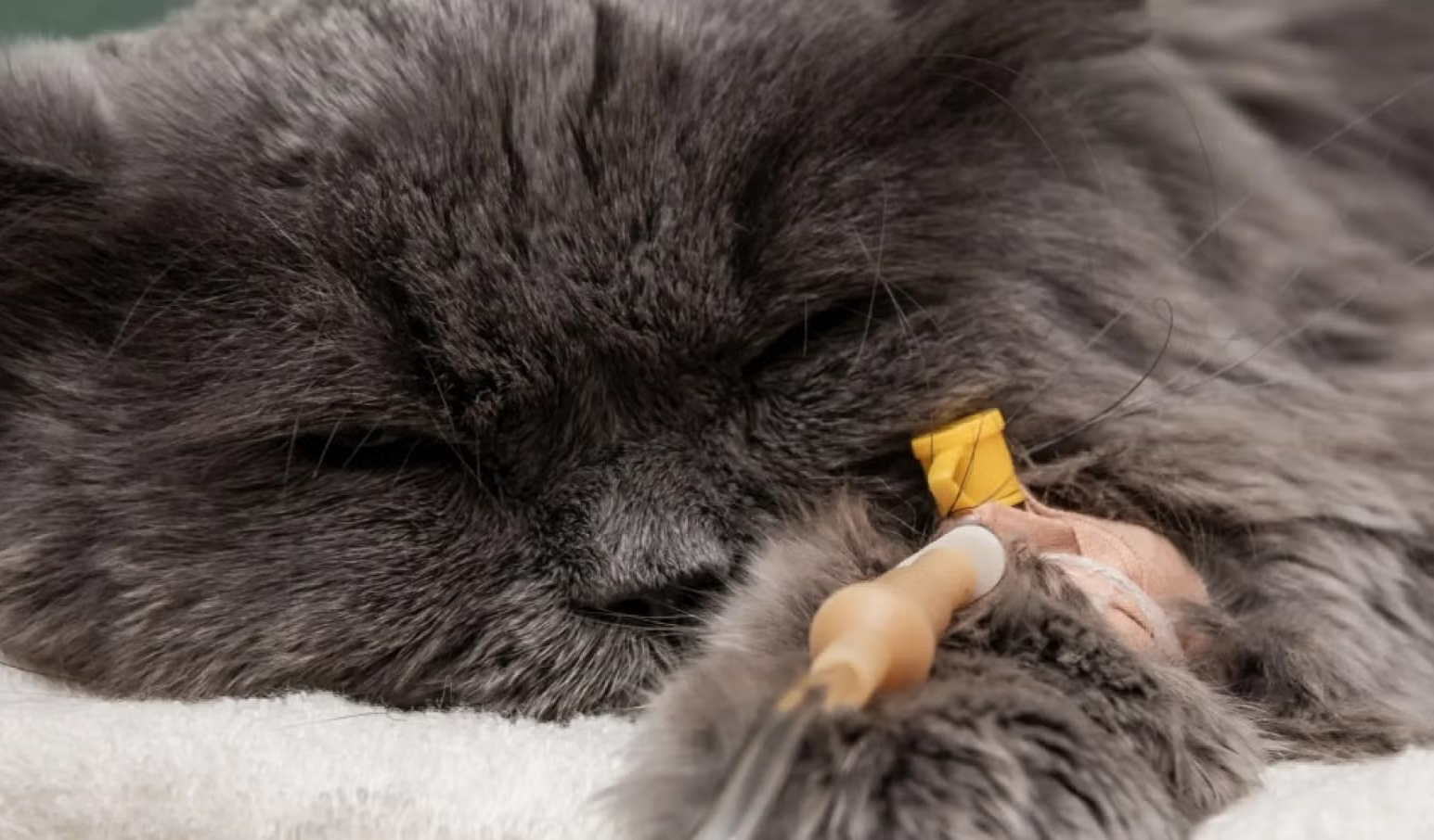
2. Chemotherapy
While chemotherapy in human medicine often causes severe side effects, the philosophy in veterinary oncology is fundamentally different. Doses and schedules are designed to control the cancer while prioritizing day-to-day comfort, minimizing risks of extreme nausea, fatigue, or infection.
                      [Common Chemotherapy Protocols]
                                     │
         ┌───────────────────────────┴───────────────────────────┐
         ▼                                                       ▼
   [COP Protocol]                                         [CHOP Protocol]
• Cyclophosphamide, Oncovin, Prednisolone             • Adds Doxorubicin (Hydroxydaunorubicin)
• Used primarily for low-grade lymphoma              • Standard for high-grade / large-cell
• Managed via oral & spaced injections                • Requires active blood monitoring
-
Managing Lymphoma: Chemotherapy is the primary treatment for systemic cancers like lymphoma. Low-grade intestinal lymphoma is managed with oral medications given at home, typically combining Chlorambucil with Prednisolone (a steroid). High-grade lymphoma requires multi-agent injectables like the CHOP or COP protocols, which rotate drugs like Cyclophosphamide, Vincristine, Doxorubicin, and Prednisolone.
-
Feline Tolerability: Cats tolerate chemotherapy remarkably well. They do not lose their body fur like humans, though they may shed their whiskers. Side effects like mild nausea or a temporary drop in white blood cells occur in less than 15% to 20% of patients and are typically managed with supportive at-home medications.
3. Radiation Therapy
Radiation oncology uses targeted high-energy beams to break the DNA bonds within cancer cells, preventing them from dividing. It is rarely used alone but is highly effective when paired with surgery. If a surgeon removes a complex tumor but the pathology report shows incomplete margins, localized radiation can target and destroy the remaining microscopic cancer cells, significantly delaying or preventing local recurrence.
Technical Summary
Successfully managing feline neoplasia depends heavily on early detection. Performing regular body checks at home during grooming or petting, scheduling routine senior veterinary exams every six months, and acting quickly at the first sign of unexplained weight loss or unusual lumps are essential steps. With an early diagnostic workup and a well-planned, personalized treatment strategy, many cats facing a neoplastic diagnosis can continue to enjoy a comfortable, active, and high-quality life for months or years to come.
                           [Clinical Oncology Summary]
                                        │
      ┌─────────────────────────────────┼─────────────────────────────────┐
      ▼                                 ▼                                 ▼
[Early Screening]               [Definitive Staging]            [Therapeutic Action]
• Bi-annual senior check-ups    • FNA / Tissue biopsy analysis  • Surgical removal with margins
• Home palpation checks         • Chest X-rays for metastasis   • Chemotherapy for lymphoma
• Monitor weight & appetite     • Abdominal ultrasound review   • Palliative pain management
FAQ
1. What is feline neoplasia?
Feline neoplasia is the abnormal and uncontrolled growth of cells in a cat's body, resulting in tumor formation. These tumors can be benign (non-cancerous) or malignant (cancerous). Malignant tumors are more common in cats than many pet owners realize and often invade surrounding tissues or spread to distant organs.
2. How common is cancer in cats?
Approximately one in five cats will develop some form of cancer during their lifetime. The risk increases significantly as cats age, making cancer one of the leading causes of death in senior felines.
3. What is the difference between benign and malignant tumors in cats?
Benign tumors remain localized and generally do not spread to other parts of the body. Malignant tumors are invasive, destroy surrounding tissues, and can metastasize through the bloodstream or lymphatic system to organs such as the lungs, liver, or lymph nodes.
4. What are the most common types of cancer in cats?
The most common feline cancers include lymphoma, squamous cell carcinoma (SCC), mammary adenocarcinoma, and feline injection-site sarcoma (FISS). Each cancer type affects different tissues and requires unique treatment approaches.
5. What is feline lymphoma?
Lymphoma is the most frequently diagnosed cancer in cats. It originates from lymphocytes, a type of white blood cell, and can develop in the gastrointestinal tract, chest cavity, kidneys, lymph nodes, or other organs.
6. What are the symptoms of feline lymphoma?
Symptoms vary depending on location but may include weight loss, vomiting, diarrhea, poor appetite, lethargy, breathing difficulties, enlarged lymph nodes, and progressive weakness.
7. What causes squamous cell carcinoma in cats?
Squamous cell carcinoma is often associated with chronic ultraviolet (UV) exposure, especially in white or lightly pigmented cats. It commonly affects the ears, nose, eyelids, and oral cavity.
8. What are the signs of oral squamous cell carcinoma?
Cats with oral SCC may develop drooling, bad breath, difficulty eating, loose teeth, facial swelling, bleeding from the mouth, and significant weight loss due to pain.
9. Why are mammary tumors so serious in cats?
Unlike dogs, over 85–90% of feline mammary tumors are malignant adenocarcinomas. They spread aggressively through lymphatic vessels and frequently metastasize to the lungs and lymph nodes.
10. Can spaying reduce the risk of mammary cancer?
Yes. Spaying before six months of age can reduce mammary cancer risk by up to 91%. Early spaying is one of the most effective preventative measures available.
11. What is feline injection-site sarcoma (FISS)?
FISS is a rare but highly aggressive cancer that develops in connective tissues at previous injection sites. It is associated with chronic inflammatory responses that may trigger malignant cellular changes.
12. What causes cancer in cats?
Cancer may result from genetic mutations, aging, viral infections such as FeLV and FIV, chronic inflammation, secondhand tobacco smoke exposure, ultraviolet radiation, and environmental carcinogens.
13. How does FeLV increase cancer risk?
Feline Leukemia Virus (FeLV) can insert viral DNA into host cells, disrupting normal cell regulation and dramatically increasing the risk of lymphoma and leukemia.
14. Does secondhand smoke affect cats?
Yes. Cats exposed to household tobacco smoke have higher rates of lymphoma and oral squamous cell carcinoma because carcinogens accumulate on their fur and are ingested during grooming.
15. How is feline cancer diagnosed?
Diagnosis typically involves physical examination, fine needle aspiration (FNA), tissue biopsy, histopathology, blood tests, X-rays, ultrasound imaging, CT scans, or MRI depending on the suspected cancer type.
16. What is a fine needle aspiration (FNA)?
FNA is a minimally invasive procedure that collects cells from a mass using a thin needle. It helps identify certain cancers but may not provide enough information for a definitive diagnosis.
17. Why is a biopsy important?
A biopsy allows veterinary pathologists to examine the tumor's structure, determine cancer type, evaluate aggressiveness, and assess whether surgical removal achieved clean margins.
18. How is feline cancer staged?
Cancer staging evaluates the extent of disease spread using imaging studies, lymph node assessment, bloodwork, and pathology results to guide treatment decisions.
19. What treatment options are available for feline cancer?
Treatment may include surgery, chemotherapy, radiation therapy, targeted therapies, immunotherapy, palliative care, or combinations of these approaches depending on the diagnosis.
20. Do cats tolerate chemotherapy well?
Most cats tolerate chemotherapy significantly better than humans. Severe side effects are uncommon, and treatment protocols focus on preserving quality of life rather than maximizing toxicity.
21. Can feline cancer be cured?
Some cancers can be cured when detected early and treated aggressively. Others can be managed for months or years through multimodal therapy, allowing cats to maintain a good quality of life.
22. What are the early warning signs of cancer in cats?
Persistent weight loss, decreased appetite, unexplained lumps, chronic wounds, difficulty breathing, vomiting, diarrhea, oral abnormalities, and changes in behavior should all prompt veterinary evaluation.
23. How often should senior cats be screened for cancer?
Cats over ten years of age should ideally undergo veterinary examinations every six months, including physical assessments and diagnostic screening when appropriate.
24. Can cancer prevention strategies reduce risk?
Yes. Early spaying, FeLV vaccination, minimizing UV exposure, avoiding tobacco smoke, maintaining a healthy weight, and regular veterinary checkups can significantly lower cancer risks.
25. What is the prognosis for cats with cancer?
Prognosis depends on cancer type, stage, location, treatment response, and overall health. Early detection generally provides the best opportunity for successful treatment and longer survival.