To the untrained eye, a small, persistent crust on a white cat’s pink ear tip or a sudden bout of drooling in an aging domestic shorthair might seem like minor concerns—perhaps a scratch from a backyard scuffle or a localized dental infection. However, in veterinary oncology, these subtle clinical presentations frequently mask one of the most aggressive, locally destructive, and painful malignancies known to feline medicine: Squamous Cell Carcinoma (SCC).
Affectionately termed “squame” by veterinary professionals, Squamous Cell Carcinoma is a malignant neoplasm derived from the differentiated squamous epithelium. This cancer accounts for approximately 15% of all cutaneous tumors in cats, making it the third most common skin malignancy behind basal cell tumors and mast cell tumors. More alarmingly, within the feline oral cavity, SCC reigns as an absolute apex predator, comprising an overwhelming 70% to 80% of all primary oral tumors.
Unlike other feline neoplasms that may present as discrete, well-encapsulated, benign masses, SCC is characterized by its sinister capacity for local infiltration, tissue necrosis, and bone destruction. While its metastatic rate to regional lymph nodes and distant visceral organs (such as the lungs) is relatively slow and late-stage compared to its rapid local destruction, the sheer anatomical location of these tumors primarily the face and the sublingual space renders them incredibly challenging to treat.
For pet parents and veterinary clinicians alike, combating feline SCC is a race against time. This human-authored, exhaustive medical masterclass will tear down complex pathological concepts, analyze the structural and environmental triggers of both cutaneous and oral SCC variations, outline cutting-edge diagnostic matrices, and detail the complete spectrum of modern therapeutic pathways required to manage this disease.
Histological Architecture and Pathophysiology of Feline SCC
To understand the aggressive nature of Squamous Cell Carcinoma, we must first examine the microscopic architecture of healthy feline skin and mucosal membranes. The epidermis and oral mucosa are composed of stratified squamous epithelium, a multi-layered cellular shield designed to protect the body against mechanical trauma, microbial invasion, and desiccation.
                  THE CELLULAR LAYERS OF THE FELINE EPIDERMIS
 ┌───────────────────────────────────────────────────────────────────────┐
 │ STRATUM CORNEUM   (Surface) - Anucleated, keratinized dead cells.     │
 ├───────────────────────────────────────────────────────────────────────┤
 │ STRATUM GRANULOSUM          - Keratohyalin granules synthesize here.  │
 ├───────────────────────────────────────────────────────────────────────┤
 │ STRATUM SPINOSUM  (Prickle) - Keratinocytes bound by desmosomes.      │
 │                                                                       │
 │ ──► ORIGIN POINT OF SQUAMOUS CELL CARCINOMA (Malicious Proliferation) │
 ├───────────────────────────────────────────────────────────────────────┤
 │ STRATUM BASALE    (Deepest) - Single layer of actively dividing cells.│
 │                                                                       │
 │ ──► Origin point of Benign Basal Cell Tumors (25% of feline skin masses)│
 └───────────────────────────────────────────────────────────────────────┘
The underlying pathophysiology of SCC involves the malignant transformation of the keratinocytes located primarily within the stratum spinosum (the prickle cell layer). Under normal conditions, these cells divide, mature, produce keratin, and gradually migrate upward to the stratum corneum before shedding.
When an oncological trigger disrupts the cellular machinery, the regulatory checks and balances governing cell division are permanently shattered. The affected squamous cells lose their structural polarity, display profound nuclear atypia (abnormal nuclei), exhibit a high mitotic index (rapid, uncontrolled division), and breach the delicate basement membrane that separates the epidermis from the underlying dermis.
Once the basement membrane is breached, the tumor transitions from an in situ (localized) state to an invasive carcinoma. The neoplastic cells secrete proteolytic enzymes, such as matrix metalloproteinases (MMPs), which systematically digest the extracellular matrix of surrounding healthy tissues. This allows the tumor to carve its way through soft tissue, muscles, nerves, and eventually, the periosteum and cortical bone of the skull or jaw.
Clinical Typologies Cutaneous vs. Oral vs. Bowenoid In Situ Carcinoma
Feline Squamous Cell Carcinoma does not manifest as a uniform disease. Instead, it presents as three distinct clinical typologies, each possessing its own unique anatomical distribution, risk factors, and prognostic pathways.
                     THE TRI-FACETED MANIFESTATION OF FELINE SCC
                                         │
         ┌───────────────────────────────┼───────────────────────────────┐
         ▼                               ▼                               ▼
  [ Cutaneous Invasive SCC ]      [ Primary Oral SCC ]            [ Bowenoid In Situ (BISC) ]
  • Sun-induced mutagenesis       • Environmentally triggered    • Multicentric presentation
  • Targets non-pigmented skin    • Highly painful; targets      • Strongly linked to FcaPV2
  • Ear pinnae, nares, eyelids    • Sublingual & gingival zones  • High risk: Devon Rex & Sphynx
1. Cutaneous Invasive Squamous Cell Carcinoma
Cutaneous invasive SCC is a classic example of environmentally induced carcinogenesis. This form of the disease is directly driven by chronic, cumulative exposure to ultraviolet (UV) radiation. It overwhelmingly targets areas of the cat’s body that lack protective coat coverage and dark melanin pigmentation.
-
Primary Predilection Sites: The absolute tips and margins of the ears (ear pinnae), the nasal planum (the hairless leathery part of the nose), the eyelids (particularly the lower margin), and the thin-haired temples just in front of the ears.
-
The Non-Pigmented Risk Factor: Cats with pure white coats, bi-color coats with white faces, or dilute coat colors (such as cream or blue) are in a state of extreme vulnerability. Melanin acts as a natural physical shield that absorbs and dissipates UV rays. Without it, the delicate skin cells receive the full, unmitigated impact of solar radiation.
2. Primary Oral Squamous Cell Carcinoma
Oral SCC is a highly malignant, agonizing disease that accounts for the vast majority of intraoral tumors in felines. Unlike its cutaneous counterpart, oral SCC is completely unlinked to solar exposure and is instead driven by a combination of chronic tissue inflammation, chemical carcinogen ingestion, and viral factors.
-
Primary Predilection Sites: The most common site is the sublingual space (directly beneath the tongue, anchoring to the lingual frenulum). It also frequently develops along the maxilla (upper gum line), the mandible (lower gum line), the tonsils, and the hard and soft palates.
-
Local Behavior: Oral SCC is incredibly aggressive locally. It exhibits a high affinity for bone invasion (osteolysis). By the time an oral tumor is visually identified by an owner, it has typically dissolved a significant portion of the underlying jawbone, leading to pathological fractures, tooth loss, and profound, unmanageable pain.
3. Bowenoid In Situ Carcinoma (BISC)
Also known as Multicentric Squamous Cell Carcinoma in Situ, Bowenoid In Situ Carcinoma (BISC) represents a unique, rare, and fascinating subset of feline cutaneous SCC, comprising roughly 10% of skin cases.
-
The In Situ Definition: In classic pathology, “in situ” denotes a cancer that is still confined to its layer of origin without breaking through the basement membrane. However, BISC is distinct because it is multicentric—meaning it develops in multiple entirely separate locations across the cat’s body simultaneously.
-
Anatomical Distribution: Unlike solar-induced SCC, BISC can develop on areas of the body that are heavily haired and entirely shielded from the sun, such as the back, shoulders, chest, and limbs.
-
Breed Predispositions: While it affects older cats of any breed, BISC displays a well-documented, severe clinical association with the Devon Rex and Sphynx breeds. In these specific cats, the disease can manifest at a significantly younger age, progress with startling speed, and break its in situ boundaries to aggressively metastasize to the internal organs, including the thoracic cavity and lungs.
Etiological Matrix Deciphering the Multifactorial Triggers
The development of Squamous Cell Carcinoma in cats is rarely a random twist of fate. Decades of epidemiological and molecular research in veterinary medicine have isolated a web of environmental, viral, and genetic triggers that drive this cellular mutation.
The Mechanisms of Solar Radiation (UVB) Mutagenesis
In cutaneous SCC, the primary driver is Ultraviolet B (UVB) radiation, which spans wavelengths between $280\text{ nm}$ and $315\text{ nm}$. When UVB light penetrates non-pigmented epidermal cells, its energy is absorbed directly by the cellular DNA.
This absorption triggers a specific chemical reaction: the formation of cyclobutane pyrimidine dimers (CPDs). These dimers cause a physical kink in the DNA double helix. If the cat’s body cannot keep up with repairing these continuous kinks, errors accumulate during cell division. Over time, this leads to permanent, irreversible mutations in critical genes that control cell growth.
                  THE PROGRESSION OF UV-INDUCED CUTANEOUS SCC
 ┌──────────────────────┐      ┌──────────────────────┐      ┌──────────────────────┐
 │ Chronic UV Exposure  │ ───► │ Solar Keratosis      │ ───► │ Invasive Cutaneous   │
 │ UVB rays damage cell │      │ Pre-cancerous zone;  │      │ Carcinoma            │
 │ DNA; kinks form in   │      │ skin becomes rough,  │      │ Basement membrane is │
 │ the double helix.    │      │ pink, and scaly.     │      │ breached by cancer.  │
 └──────────────────────┘      └──────────────────────┘      └──────────────────────┘
The primary target of this solar damage is the p53 tumor suppressor gene. Often called the “guardian of the genome,” the p53 gene produces a protein that halts cell division if DNA damage is detected, allowing the cell time to repair itself, or triggering cell suicide (apoptosis) if the damage is too severe. When UVB radiation mutates and knocks out the p53 gene, the damaged epidermal cells are free to replicate indefinitely, culminating in invasive cutaneous SCC.
The Viral Element: Feline Papillomavirus (FcaPV)
While solar damage dominates the cutaneous landscape, viral pathogens play an equally critical role, particularly in Bowenoid In Situ Carcinoma (BISC) and a significant portion of oral and cutaneous cases.
Veterinary PCR testing has revealed that between 25% and 33% of feline skin SCC lesions harbor Feline Papillomavirus DNA.
-
FcaPV2 (Feline Papillomavirus Type 2): This is the primary viral driver isolated in feline SCC. It produces specific viral oncoproteins (designated as E6 and E7) that bind to and deactivate the cat’s natural tumor suppressor proteins (p53 and Retinoblastoma protein, or pRb).
-
Other Strains: FcaPV3, 4, 5, and 6 have also been detected, though less frequently. These viruses can hide silently in the skin of healthy cats, but when combined with UV damage or immune suppression, they act as an accelerator for cancer development.
The Environmental Hazards of Oral Carcinogenesis
The intraoral cavity of a cat is highly vulnerable to chemical toxins due to their meticulous grooming behavior. When carcinogens settle out of the household air onto a cat’s fur coat, the cat acts as a biological vacuum cleaner, licking up and ingesting these compounds during daily self-grooming.
                       ORAL CHEMICAL INGESTION PATHWAY
 ┌────────────────────────┐      ┌────────────────────────┐      ┌────────────────────────┐
 │ Carcinogens in Air     │ ───► │ Grooming Accumulation  │ ───► │ Direct Tissue Impact   │
 │ Tobacco smoke, flea    │      │ Toxins blanket the fur │      │ Licking concentrates   │
 │ collar residues settle.│      │ coat of the cat.       │      │ carcinogens on mucosa. │
 └────────────────────────┘      └────────────────────────┘      └────────────────────────┘
Epidemiological data has identified several home environmental factors that dramatically increase the statistical risk of oral SCC:
-
Environmental Tobacco Smoke (ETS): Cats living in smoking environments face a 4.5x higher risk of developing oral SCC. The airborne tar, nicotine, and heavy metals bind to the fur, which the cat then deposits directly onto the sensitive mucosal tissues beneath the tongue during grooming.
-
Flea Collars (5.3x Increased Risk): Older generations of chemical flea collars continuously emit volatile pesticide residues across the neck and facial fur. As cats groom their faces and forelimbs, they experience prolonged, concentrated contact with these potential carcinogens.
-
Canned Tuna (4.7x Increased Risk) & Industrial Canned Diets (3.6x Increased Risk): Regular consumption of commercial canned food diets—and canned tuna specifically—is statistically linked to oral SCC. While wet food provides excellent hydration benefits, the physical action of eating wet food allows food particles to stick to the gums and under the tongue for extended periods. This can lead to chronic, low-grade tissue inflammation, which may act as a catalyst for cellular mutations over time.
Systemic Immune Suppression (FeLV and FIV)
Cats infected with Feline Leukemia Virus (FeLV) or Feline Immunodeficiency Virus (FIV) are at a significantly higher risk for various cancers, including aggressive SCC. These retroviruses attack and weaken the cat’s cellular immune system.
A healthy immune system relies on “immune surveillance” to constantly identify and destroy abnormal, mutated cells before they can form a solid tumor. When FeLV or FIV compromises this system, early cancer cells can grow unchecked, bypassing the body’s natural defenses.
Clinical Presentations and Diagnostic Red Flags
Early detection is the single most important factor determining whether a cat survives Squamous Cell Carcinoma. Unfortunately, because early SCC easily mimics benign skin and dental conditions, many cases are caught far too late.
Cutaneous SCC Symptoms: The Illusion of the Healing Wound
Cutaneous SCC rarely starts as a noticeable, raised lump. Instead, it begins as a subtle lesion called solar keratosis. At this stage, the skin looks slightly red, rough, and scaly, and the hair may begin to thin.
As the cancer breaches the basement membrane and becomes invasive, the clinical presentation shifts:
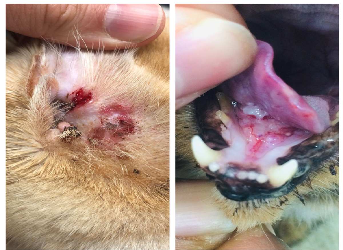
                   CUTANEOUS INVASIVE SCC VISUAL PROFILE
  Lesion Presentation         Visual and Tactile Characteristics
  ────────────────────────────────────────────────────────────────────────
  Ulcerated Fissures   ──► Deep, raw cracks in the nasal planum that bleed easily.
  Adherent Crusts      ──► Thick, dark scabs on the ear margins that keep peeling off.
  Erosive Crater       ──► Sunken, open sores on the eyelids that gradually erode tissue.
The most critical diagnostic red flag for a cat owner or clinician is a scab or sore on the face or ears that fails to heal after two weeks of standard medical therapy.
The Steroid/Antibiotic Diagnostic Test
When a cat presents with an irritated, crusty facial sore, initial treatments often target common issues like a bacterial skin infection (pyoderma), feline acne, or an allergic reaction (atopic dermatitis). The vet will typically prescribe a course of antibiotics combined with a topical or systemic steroid like prednisolone.
If the lesion is a benign infection or allergy, it will show dramatic, visible healing within 7 to 14 days. An SCC lesion will not improve; instead, it will hold its ground or grow larger, angrier, and more ulcerated. Any facial lesion that fails this therapeutic trial requires immediate diagnostic biopsy.
Oral SCC Symptoms: The Silent Agony
Oral SCC is notoriously difficult to spot early on. Cats are biologically hardwired to hide chronic pain, an evolutionary trait designed to prevent them from looking vulnerable to predators. Consequently, by the time a cat shows clear signs of mouth pain, the intraoral tumor is usually quite advanced.
Owners should watch for these critical behavioral and physical warning signs:
-
Inexplicable Weight Loss and Inappetence: The cat approaches the food bowl, clearly hungry, but sniffs the food and hesitates or walks away.
-
Behavioral Aggression Toward Food: The cat may hiss, growl, or swat at the food bowl. This happens because they associate the physical act of eating with sudden, sharp pain.
-
Hypersalivation (Drooling): The cat produces an excess of thick, ropy saliva. As the tumor ulcerates and tissue dies (necrosis), this drooling often turns pinkish or bloody and develops a foul, putrid odor (halitosis).
-
Pawing at the Face: The cat frequently rubs their mouth against furniture or frantically paws at their jaw as if trying to dislodge a foreign object.
-
Asymmetry of the Face and Mouth: Looking at the cat dead-on may reveal a swollen jaw, a bulging cheek, or an eye that looks slightly pushed out of place. Inside the mouth, a large sublingual mass may physically push the tongue to one side, preventing the cat from drawing it back into their mouth.
                   ORAL SCC MISDIAGNOSIS PITFALL MATRIX
  True Oncological Condition      Common Clinical Misdiagnosis
  ────────────────────────────────────────────────────────────────────────
  Alveolar Ridge Gum SCC     ──► Severe Periodontal Disease / Gingivitis
  Jaw Bone Invasion / Lysis  ──► Tooth Root Abscess / Facial Swelling
  Erosive Palate Lesion      ──► Feline Eosinophilic Ulcer / Rodent Ulcer
Because an oral tumor can look strikingly similar to severe dental disease or a tooth root abscess, a sedated, thorough oral examination combined with diagnostic imaging is essential to avoid treating a malignant tumor as a simple dental issue.
Bowenoid In Situ Carcinoma (BISC) Symptoms: The Multicentric Patches
BISC presents with a completely different clinical look than classic cutaneous SCC. Instead of a single, raw, bleeding ulcer on the ear or nose, BISC shows up as multiple, clearly defined, flat patches or raised bumps across the body.
These lesions are typically dark brown or black, rough, and covered in a thick crust. They are often found on the neck, shoulders, chest, and belly. Because they look like harmless pigment changes or old scabs, they can easily be mistaken for flea allergy dermatitis or superficial infections until they begin to spread across wider areas of the skin.
The Diagnostic Protocol: Biopsy, Cytology, and Staging
When a clinical exam raises suspicion of Squamous Cell Carcinoma, confirming the diagnosis and determining the stage of the disease requires a structured sequence of diagnostic tests.
                  DIAGNOSTIC WORKFLOW FOR SUSPECTED FELINE SCC
 ┌───────────────────────────┼───────────────────────────┐
 │                           │                           │
 ▼                           ▼                           ▼
[ Punch/Incisional Biopsy ] [ Fine Needle Aspirate ]    [ Advanced Imaging ]
The definitive test.        Used to evaluate regional   3-view chest X-rays or CT
Histopathology confirms     lymph nodes for early       scans to rule out internal
malignancy & growth style.  metastatic spread.          metastasis and bone damage.
1. Histopathology: The Definitive Biopsy
While a doctor may strongly suspect SCC based on a lesion’s appearance, a tissue biopsy remains the gold standard for a definitive diagnosis.
-
Why Cytology (FNA) Often Fails Initially: Attempting a fine needle aspirate (FNA) on a raw, open SCC sore often yields disappointing results. The surface of these tumors is typically blanketed by a thick layer of inflammatory cells, dead tissue, and secondary bacteria. A needle scan of this surface layer often shows only “severe inflammation,” completely missing the malignant cancer cells buried underneath.
-
Biopsy Techniques: For skin lesions, a veterinarian will use a specialized circular blade called a punch biopsy to harvest a small, full-thickness cylinder of tissue from the edge of the sore, ensuring they capture both abnormal and healthy tissue. For oral masses, an incisional biopsy is performed under deep sedation or general anesthesia, taking care not to disrupt critical blood vessels or nerves.
-
What the Pathologist Sees: Under the microscope, a veterinary pathologist looks for characteristic signs of malignant squamous proliferation: cords and sheets of atypical epithelial cells driving deep into the dermis, high numbers of abnormal cell divisions (mitotic figures), individual cells dying prematurely (dyskeratosis), and the formation of keratin pearls—concentric nests of laminated keratin pushed inside the tumor tissue as the cells attempt to mature abnormally.
2. Clinical Staging: Assessing the Reach of the Disease
Once histopathology confirms the presence of SCC, the next critical step is to determine if and where the cancer has spread. This process, known as clinical staging, guides the entire treatment strategy and shapes the cat’s long-term prognosis.
-
Lymph Node Evaluation: The vet will perform an FNA on the regional lymph nodes that drain the head and neck—specifically the submandibular and prescapular lymph nodes. These samples are carefully evaluated under a microscope to check for early signs of migrating cancer cells.
-
Three-View Thoracic Radiographs: Even though SCC is slow to spread internally, taking three distinct views of the chest (left lateral, right lateral, and ventrodorsal) is essential to ensure the cancer hasn’t metastasized to the lungs.
-
Advanced Computed Tomography (CT Scans): For oral SCC and advanced cutaneous cases on the skull, a CT scan is the premier choice for imaging. Standard X-rays can easily miss early bone damage. A high-resolution CT scan provides a detailed, three-dimensional view of the skull, showing exactly how much of the jaw or facial bones have been invaded by the tumor. This mapping is vital if the veterinary surgeon hopes to plan a successful surgical removal.
Comprehensive Therapeutic Modalities
Managing feline Squamous Cell Carcinoma requires an aggressive, multi-layered treatment plan. Because this cancer is highly invasive locally, treating it successfully demands a combination of surgery, advanced technology, and supportive care.
Deep Dive into Surgical Interventions
Surgery remains the single most reliable weapon against Squamous Cell Carcinoma, provided the tumor is caught early and sits in an area where a surgeon can safely cut wide boundaries around it.
1. Pinnectomy (Amputation of the Ear Flap)
When SCC targets the margins of the ears, a pinnectomy is the standard treatment of choice. The surgeon amputates the affected portion of the outer ear flap, cutting well below the visible margins of the tumor to ensure no microscopic cancer cells are left behind.
                  PINNECTOMY SURGICAL MARGIN PROTOCOL
  [ Healthy Ear Base ] ───────► [ Clean $2\text{ cm}$ Border ] ───────► [ Malignant Tumor Margin ]
  (Preserved tissue)            (Surgical cut line)             (Ulcerated ear tip)
While losing an ear flap changes a cat’s appearance, giving them a unique, asymmetrical look, it is an exceptionally well-tolerated surgery. Cats heal quickly, experience immediate relief from the itchy, painful sore, and can go on to live a completely normal, cancer-free life if the surgical boundaries are clear.
2. Mandibulectomy and Maxilectomy
For oral SCC that is caught early and confined to the gum line of the upper or lower jaw, a board-certified veterinary surgeon can perform a partial mandibulectomy (removing part of the lower jaw bone) or a partial maxillectomy (removing part of the upper jaw bone).
Because feline oral SCC aggressively invades bone, simply scraping the tumor off the surface is a guaranteed recipe for rapid failure. The surgeon must cut directly through the healthy bone surrounding the tumor to achieve a true cure.
While this sounds like a drastic step, cats are incredibly resilient. With dedicated post-operative care, specialized liquid nutrition, and proper pain management, most cats adapt remarkably well to their altered jaw structure, learning how to scoop up canned food within a few weeks.
The Tragedy of Sublingual Oral SCC
Unfortunately, the prognosis changes drastically if the oral SCC develops in the sublingual space beneath the tongue. This area is rich in vital blood vessels (the lingual arteries) and critical nerves, and it anchors the tongue muscles needed for lapping water and swallowing food.
Surgically removing a tumor in this location would require amputating a large portion of the tongue itself. Cats cannot survive or maintain a reasonable quality of life without a functional tongue, as they become permanently unable to eat, drink, or groom. For this reason, surgical removal of sublingual SCC is rarely recommended by veterinary oncologists, as it causes immense post-operative suffering without offering a meaningful cure.
Palliative Care Protocols and Advanced Quality of Life Management
When a diagnosis of advanced, non-resectable oral SCC is delivered, the clinical focus shifts completely from seeking a cure to maximizing comfort and maintaining the cat’s quality of life for as long as possible.
The Multi-Modal Pain Management Protocol
Advanced oral SCC causes severe, ongoing pain due to both bone destruction and constant tissue inflammation. Managing this discomfort requires a multi-layered pain plan designed by a veterinarian:
-
Non-Steroidal Anti-Inflammatory Drugs (NSAIDs): Medications such as Meloxicam or Piroxicam are frequently used. Beyond providing basic pain relief, these specific drugs work as cyclooxygenase (COX) inhibitors. Many feline oral SCC tumors overexpress the COX enzyme, which drives tumor inflammation and helps the cancer grow new blood vessels. By blocking this enzyme, NSAIDs can actually help slow down the tumor’s growth while easing inflammation. However, because these medications are processed by the kidneys, they must be used with caution, requiring regular blood tests to safeguard the cat against renal failure.
-
Targeted Opioid Therapy (Buprenorphine): For immediate, reliable pain relief, Buprenorphine is a standout choice in feline medicine. This medication can be administered directly onto the cat’s oral mucous membranes (transmucosal), where it is absorbed straight into the bloodstream without requiring the cat to swallow a pill. This makes it an incredibly low-stress way to deliver powerful pain management to a cat with a sore mouth.
-
Nerve Pain Management (Gabapentin): As the oral tumor grows and begins to press against the major nerves of the face and jaw, it can trigger sharp, burning nerve pain. Adding Gabapentin to the cat’s routine helps soothe these overactive pain signals, working hand-in-hand with NSAIDs and opioids to keep the cat calm and comfortable.
The Role of “Magic Mouthwash” in End-of-Life Support
To help cats with advanced oral tumors continue to enjoy their food, veterinarians often prescribe a specialized local pain-relieving compound known as Magic Mouthwash.
This oral solution combines an anesthetic, an antihistamine, and an antacid in equal measures:
How It Works:
-
2% Viscous Lidocaine: Acts as a powerful local anesthetic, temporarily numbing the raw, ulcerated surfaces of the tumor on contact.
-
Liquid Diphenhydramine (Benadryl): Works locally to reduce swelling, ease itching, and provide a mild, calming effect.
-
Maalox (Aluminum and Magnesium Hydroxide): Coats the sensitive lining of the mouth, helping the active pain-relieving ingredients stick to the tumor surface longer while neutralizing irritating oral acids.
Administration Guide:
The owner gently applies a small, precise dose of this mixture directly into the side of the cat’s mouth using a needleless syringe roughly 30 minutes before feeding. This temporarily numbs the raw sores, allowing the cat to eat a meal of soft, blended food without experiencing sharp pain.
⚠️ Important Clinical Warning: This mouthwash is a short-term, superficial measure designed purely for comfort care. It provides temporary relief from surface pain, but it does not stop or slow down the underlying spread of the cancer.
Modifying the Diet for Oral Comfort
When a cat has an oral tumor, their food must be modified to make swallowing as effortless as possible. Standard dry kibble should be completely removed from the diet, as the hard edges can scrape against the tumor, causing pain and bleeding.
Instead, switch to high-calorie, highly palatable wet foods. Blend the canned food with warm water or low-sodium bone broth until it reaches a completely smooth, liquid consistency, often called a gruel.
                  OPTIMAL LIQUID DIET PROFILE FOR ORAL SCC
 ┌───────────────────────────────────────────────────────────────────────┐
 │ SMOOTH CONSISTENCY - Blended into a liquid gruel; eliminates chewing. │
 ├───────────────────────────────────────────────────────────────────────┐
 │ ELEVATED BOWLS     - Placed on a raised stand; allows gravity to     │
 │                      help the cat swallow food with less tongue movement.│
 ├───────────────────────────────────────────────────────────────────────┐
 │ WARMED TEMPERATURE - Served warm to boost the food's aroma, coaxing   │
 │                      a cat whose sense of smell may be muted by illness.│
 └───────────────────────────────────────────────────────────────────────┘
Serving the food in wide, flat, shallow dishes or on a slightly raised stand allows the cat to lap up the liquid easily, using gravity to help them swallow with minimal tongue movement.
Proactive Prevention and Long-Term Environmental Protection
While treating an established case of Squamous Cell Carcinoma is a major medical undertaking, preventing the disease from taking hold is entirely achievable through thoughtful, proactive changes to a cat’s lifestyle and home environment.
1. Advanced Sun Safety and Home UV Protection
If you share your home with a white, light-colored, or orange tabby cat, managing their sun exposure is your first line of defense against cutaneous SCC.
-
Enforce Smart Indoor Hours: Keep high-risk cats indoors during peak sunlight hours, typically between 10:00 AM and 2:00 PM, when the sun’s UVB rays are at their strongest and most damaging.
-
Install UV Window Protection: Many indoor cats love to spend hours basking in sunny windows. Standard window glass blocks UVB rays, but it allows UVA rays to pass right through, which can still contribute to long-term skin damage. Installing clear, specialized UV-blocking window films on your home’s sunniest windows filters out up to 99% of harmful radiation. This allows your cat to enjoy the warmth of the sun without any of the associated cancer risks.
-
Use Pet-Safe Sunscreen: If your light-colored cat has outdoor access, you can apply a thin layer of specialized sunscreen to the tips of their ears and the bridge of their nose.
🚫 Critical Ingredient Warning: Never use human sunscreens on a cat. Human products frequently contain Zinc Oxide or Salicylates (Aspirin derivatives). Because cats groom themselves constantly, they will quickly lick the sunscreen off their skin. Ingesting zinc oxide can cause severe, life-threatening red blood cell destruction (hemolytic anemia) in felines, while salicylates can trigger acute liver and kidney failure. Always choose a product that is explicitly labeled as non-toxic and safe for cats.
2. Cleaning Up the Indoor Environment
Given the clear, proven link between environmental toxins and oral SCC, removing common chemical hazards from your home is a vital preventative step.
-
Establish a Strict Smoke-Free Home: If you smoke or use tobacco products, always step outside away from your pets, or consider quitting entirely. Eliminating secondhand smoke removes the single most dangerous source of airborne carcinogens that settle on your cat’s coat and end up in their mouth during grooming.
-
Ditch Old-Fashioned Flea Collars: Avoid using older, dust-generating chemical flea collars. Instead, work with your veterinarian to choose modern, safe, and highly effective flea preventatives, such as topical spot-on treatments or oral medications that control pests without leaving hazardous residues on your cat’s fur.
-
Offer a Diverse, Balanced Diet: While wet food provides undeniable health benefits for a cat’s hydration and kidney function, avoid feeding a single protein source—like canned tuna—exclusively for years on end. Alternating between high-quality wet foods and crisp, clean kibble can help reduce the chronic, low-grade gum inflammation associated with soft diets while keeping your cat’s nutrition balanced.
3. Immunological Support Through Life-Stage Nutrition
A resilient, highly active immune system is a cat’s best internal defense against the viral pathways that can accelerate cancer growth, such as Feline Papillomavirus. Ensuring your cat receives targeted, premium nutrition during critical stages of life can help build a strong foundation for long-term health.
                  CELLULAR SUPPORT SYSTEM VIA NUTRITION
 ┌───────────────────────────────────────────────────────────────────────┐
 │ BOVINE COLOSTRUM   - Delivers raw immunoglobulins to strengthen the  │
 │                      body's natural defense against viral pathogens. │
 ├───────────────────────────────────────────────────────────────────────┐
 │ OMEGA-3 FATTY ACIDS - High levels of DHA and EPA work to reduce       │
 │                       chronic, low-grade tissue inflammation.         │
 └───────────────────────────────────────────────────────────────────────┘
When selecting a diet for growing kittens or young adults, look for premium formulas fortified with bovine colostrum (such as Pro Plan Kitten Starter). Colostrum is rich in natural antibodies and bioactive proteins that help bridge early immunity gaps, helping the body fight off early viral threats.
Additionally, ensuring your cat’s adult diet contains robust levels of Omega-3 fatty acids (DHA and EPA) from high-quality marine sources helps keep systemic inflammation in check, protecting delicate mucosal tissues and supporting overall cellular health as they age.
Final Clinical Outlook: The Power of Watchful Care
Feline Squamous Cell Carcinoma remains a formidable and deeply serious diagnosis in veterinary medicine. For cats facing an advanced sublingual oral tumor, the journey is often brief, focusing on providing a gentle, pain-free end-of-life chapter through dedicated palliative care.
Yet, for cutaneous and early-stage skin variations, the story can have a completely different, triumphant ending. When a pet parent catches the warning signs early—spotting that tiny, unhealing ear scab or rough patch of skin—and pursues a swift diagnosis and wide surgical removal, the prognosis shifts from dark to bright.
By taking simple, proactive steps to safeguard your home—shielding your cat from peak UV rays, choosing safe, low-dust products, keeping indoor air free of smoke, and supporting their body with life-stage nutrition—you can protect your feline companion from this silent threat, ensuring they enjoy a long, comfortable, and healthy life by your side.
FAQ – Feline Squamous Cell Carcinoma (SCC)
1. What is Squamous Cell Carcinoma (SCC) in cats?
Squamous Cell Carcinoma (SCC) is a malignant cancer that develops from squamous epithelial cells found in the skin, mouth, nose, ears, and mucosal tissues. It is one of the most aggressive and painful cancers in cats, especially when it affects the oral cavity.
2. What causes SCC in cats?
Several factors can trigger SCC in cats, including:
- Chronic UV/sun exposure
- White or light-colored fur with low pigmentation
- Environmental tobacco smoke
- Feline Papillomavirus (FcaPV)
- Chronic oral inflammation
- Chemical exposure from flea collars or toxins
- Immune suppression from FeLV or FIV
3. Which cats are most at risk for SCC?
Cats at higher risk include:
- White cats or cats with pink noses and ears
- Outdoor cats with prolonged sun exposure
- Older cats (typically over 10 years old)
- Cats exposed to cigarette smoke
- Devon Rex and Sphynx breeds (higher BISC risk)
4. What are the early signs of SCC in cats?
Common early warning signs include:
- Crusty sores on the ears or nose
- Non-healing scabs
- Red or ulcerated skin patches
- Drooling or bad breath
- Difficulty eating
- Weight loss
- Loose teeth
- Swelling around the jaw or mouth
5. How do I know if a sore is cancerous?
A lesion that does not heal within 2 weeks despite antibiotics or anti-inflammatory treatment should be evaluated immediately by a veterinarian. Persistent sores are a major red flag for SCC.
6. Is SCC in cats painful?
Yes. SCC is extremely painful, especially oral SCC. As the tumor invades soft tissue and bone, cats may experience severe pain while eating, grooming, or swallowing.
7. Can SCC spread to other organs?
SCC primarily causes aggressive local tissue destruction first. It can eventually spread to lymph nodes and lungs, but metastasis usually occurs later in the disease progression.
8. How is SCC diagnosed in cats?
Veterinarians typically diagnose SCC using:
- Physical examination
- Tissue biopsy
- Histopathology
- Fine needle aspiration (FNA)
- X-rays or CT scans
- Lymph node evaluation
A biopsy is the definitive diagnostic method.
9. What does oral SCC look like in cats?
Oral SCC may appear as:
- A mass under the tongue
- Bleeding gums
- Swollen jaw
- Loose teeth
- Ulcerated tissue
- Excessive drooling
- Foul mouth odor
Many cases resemble severe dental disease.
10. Can SCC in cats be cured?
Early-stage cutaneous SCC can sometimes be cured with aggressive surgery. Advanced oral SCC is much harder to cure because tumors often invade bone before detection.
11. What treatments are available for feline SCC?
Treatment options include:
- Surgical removal
- Pinnectomy (ear tip amputation)
- Partial jaw surgery
- Radiation therapy
- Cryotherapy
- Photodynamic therapy
- NSAIDs
- Pain management medications
- Palliative care
12. What is the survival rate for cats with SCC?
Survival depends heavily on tumor location and stage:
- Early ear/nose SCC: Often excellent prognosis after surgery
- Advanced oral SCC: Typically poor prognosis
- Untreated SCC: Progresses rapidly and painfully
Early detection dramatically improves outcomes.
13. Can indoor cats develop SCC?
Yes. Indoor cats can still develop SCC due to:
- Window UV exposure
- Viral infections
- Chronic inflammation
- Environmental toxins
- Genetic predisposition
14. Is SCC contagious between cats?
No. The cancer itself is not contagious. However, some contributing viral factors, such as Feline Papillomavirus, may spread between cats.
15. Can sunlight really cause skin cancer in cats?
Yes. Chronic UVB exposure damages DNA in skin cells, especially in white or lightly pigmented cats. This is one of the leading causes of cutaneous SCC.
16. How can I prevent SCC in my cat?
Preventive measures include:
- Limiting sun exposure
- Keeping white cats indoors during peak sunlight hours
- Using pet-safe sunscreen
- Avoiding cigarette smoke exposure
- Using safer flea preventatives
- Maintaining dental hygiene
- Providing high-quality nutrition
17. What is Bowenoid In Situ Carcinoma (BISC)?
BISC is a rare multicentric form of SCC linked to Feline Papillomavirus. It appears as multiple dark crusted skin lesions across the body and is more common in Devon Rex and Sphynx cats.
18. Why does oral SCC cause drooling?
Tumors inside the mouth irritate tissues, create ulcers, and interfere with swallowing. This leads to excessive saliva production, often mixed with blood or pus.
19. Can cats still eat with oral SCC?
Many cats struggle to eat due to severe pain. Soft blended foods, liquid diets, pain management, and supportive care can help maintain comfort and nutrition.
20. When should I take my cat to the vet?
Seek immediate veterinary care if your cat has:
- A sore that will not heal
- Persistent drooling
- Facial swelling
- Bleeding from the mouth
- Difficulty eating
- Weight loss
- Bad breath
- Eye or nose ulcers
- Chronic ear crusting
Early diagnosis is critical for improving survival and comfort.
 (1)")